


The Role of Phosphate Binding in Purine Nucleoside Phosphorylase of Helicobacter pylori
Bosnjakovic, M., Lescic Asler, I., Stefanic, Z.(2018) Croat Chem Acta 91
Experimental Data Snapshot
Starting Model: experimental
View more details
wwPDB Validation 3D Report Full Report

(2018) Croat Chem Acta 91
Entity ID: 1 | |||||
|---|---|---|---|---|---|
| Molecule | Chains | Sequence Length | Organism | Details | Image |
| Purine nucleoside phosphorylase DeoD-type | 233 | Helicobacter pylori 26695 | Mutation(s): 0 Gene Names: deoD, HP_1178 EC: 2.4.2.1 | 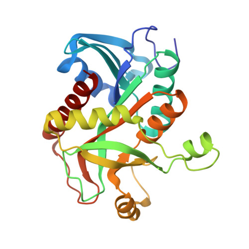 | |
UniProt | |||||
Entity Groups | |||||
| Sequence Clusters | 30% Identity50% Identity70% Identity90% Identity95% Identity100% Identity | ||||
| UniProt Group | P56463 | ||||
Sequence AnnotationsExpand | |||||
Reference Sequence | |||||
| Ligands 2 Unique | |||||
|---|---|---|---|---|---|
| ID | Chains | Name / Formula / InChI Key | 2D Diagram | 3D Interactions | |
| PO4 Download:Ideal Coordinates CCD File | DA [auth E] IA [auth F] J [auth A] P [auth B] U [auth C] | PHOSPHATE ION O4 P NBIIXXVUZAFLBC-UHFFFAOYSA-K |  | ||
| IMD Download:Ideal Coordinates CCD File | AA [auth E] BA [auth E] CA [auth E] EA [auth F] FA [auth F] | IMIDAZOLE C3 H5 N2 RAXXELZNTBOGNW-UHFFFAOYSA-O |  | ||
| Length ( Å ) | Angle ( ˚ ) |
|---|---|
| a = 93.32 | α = 90 |
| b = 91.49 | β = 119.9 |
| c = 93.41 | γ = 90 |
| Software Name | Purpose |
|---|---|
| PHENIX | refinement |
| SCALA | data scaling |
| PDB_EXTRACT | data extraction |
| MOSFLM | data reduction |
| MOLREP | phasing |
| Funding Organization | Location | Grant Number |
|---|---|---|
| Croatia | 7423 |


