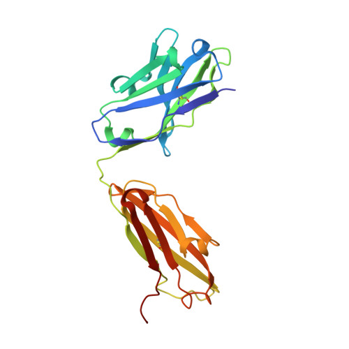
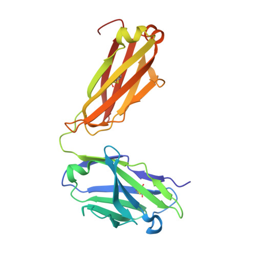
6H3H

Fab fragment of antibody against fullerene C60


Experimental Data Snapshot

  • Method: X-RAY DIFFRACTION
  • Resolution: 1.92 Å
  • R-Value Free: 0.234 
  • R-Value Work: 0.188 
  • R-Value Observed: 0.191 

Starting Model: experimental
View more details

wwPDB Validation   3D Report Full Report


This is version 1.2 of the entry. See complete history


Literature

Structure of the Anti-C60 Fullerene Antibody Fab Fragment: Structural Determinants of Fullerene Binding.

Osipov, E.M.Hendrickson, O.D.Tikhonova, T.V.Zherdev, A.V.Solopova, O.N.Sveshnikov, P.G.Dzantiev, B.B.Popov, V.O.

(2019) Acta Naturae 11: 58-65

  • Primary Citation of Related Structures:  
    6H3H

  • PubMed Abstract: 

    The structure of the anti-C 60 fullerene antibody Fab fragment (FabC 60 ) was solved by X-ray crystallography. The computer-aided docking of C 60 into the antigen-binding pocket of FabC 60 showed that binding of C 60 to FabC 60 is governed by the enthalpy and entropy; namely, by π-π stacking interactions with aromatic residues of the antigen-binding site and reduction of the solvent-accessible area of the hydrophobic surface of C 60 . A fragment of the mobile CDR H3 loop located on the surface of FabC 60 interferes with C 60 binding in the antigen-binding site, thereby resulting in low antibody affinity for C 60 . The structure of apo-FabC 60 has been deposited with pdbid 6H3H.


  • Organizational Affiliation

    Bach Institute of Biochemistry, Research Center of Biotechnology of the Russian Academy of Sciences, Leninsky Ave. 33, 119071, Moscow, Russia.


Macromolecules
Find similar proteins by:  (by identity cutoff)  |  3D Structure
Entity ID: 1
MoleculeChains Sequence LengthOrganismDetailsImage
Anti-fullerene antibody Fab fragment Heavy chain
A, C
222Mus musculusMutation(s): 0 
Entity Groups  
Sequence Clusters30% Identity50% Identity70% Identity90% Identity95% Identity100% Identity
Sequence Annotations
Expand
  • Reference Sequence
Find similar proteins by:  (by identity cutoff)  |  3D Structure
Entity ID: 2
MoleculeChains Sequence LengthOrganismDetailsImage
Anti-fullerene antibody Fab fragment Light chain
B, D
215Mus musculusMutation(s): 0 
Entity Groups  
Sequence Clusters30% Identity50% Identity70% Identity90% Identity95% Identity100% Identity
Sequence Annotations
Expand
  • Reference Sequence
Small Molecules
Ligands 2 Unique
IDChains Name / Formula / InChI Key2D Diagram3D Interactions
SO4
Query on SO4

Download Ideal Coordinates CCD File 
G [auth B]
H [auth B]
I [auth C]
L [auth D]
M [auth D]
G [auth B],
H [auth B],
I [auth C],
L [auth D],
M [auth D],
N [auth D]
SULFATE ION
O4 S
QAOWNCQODCNURD-UHFFFAOYSA-L
GOL
Query on GOL

Download Ideal Coordinates CCD File 
E [auth A],
F [auth B],
J [auth D],
K [auth D]
GLYCEROL
C3 H8 O3
PEDCQBHIVMGVHV-UHFFFAOYSA-N
Experimental Data & Validation

Experimental Data

  • Method: X-RAY DIFFRACTION
  • Resolution: 1.92 Å
  • R-Value Free: 0.234 
  • R-Value Work: 0.188 
  • R-Value Observed: 0.191 
  • Space Group: P 1 21 1
Unit Cell:
Length ( Å )Angle ( ˚ )
a = 40.33α = 90
b = 138.18β = 91.85
c = 83.53γ = 90
Software Package:
Software NamePurpose
REFMACrefinement
XDSdata reduction
XSCALEdata scaling
BALBESphasing

Structure Validation

View Full Validation Report


Entry History & Funding Information

Deposition Data


Funding OrganizationLocationGrant Number
Russian Science FoundationRussian Federation14-24-00172

Revision History  (Full details and data files)

  • Version 1.0: 2019-05-15
    Type: Initial release
  • Version 1.1: 2024-01-17
    Changes: Data collection, Database references, Refinement description
  • Version 1.2: 2024-11-06
    Changes: Structure summary