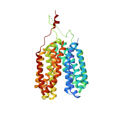
6HCL

Crystal structure of a MFS transporter with Ligand at 2.69 Angstroem resolution


Experimental Data Snapshot

  • Method: X-RAY DIFFRACTION
  • Resolution: 2.50 Å
  • R-Value Free: 0.259 
  • R-Value Work: 0.239 
  • R-Value Observed: 0.240 

Starting Model: experimental
View more details

wwPDB Validation   3D Report Full Report


Ligand Structure Quality Assessment 


This is version 1.2 of the entry. See complete history


Literature

Mechanistic basis of L-lactate transport in the SLC16 solute carrier family.

Bosshart, P.D.Kalbermatter, D.Bonetti, S.Fotiadis, D.

(2019) Nat Commun 10: 2649-2649

  • DOI: https://doi.org/10.1038/s41467-019-10566-6
  • Primary Citation of Related Structures:  
    6G9X, 6HCL

  • PubMed Abstract: 

    In human and other mammalian cells, transport of L-lactate across plasma membranes is mainly catalyzed by monocarboxylate transporters (MCTs) of the SLC16 solute carrier family. MCTs play an important role in cancer metabolism and are promising targets for tumor treatment. Here, we report the crystal structures of an SLC16 family homologue with two different bound ligands at 2.54 and 2.69 Å resolution. The structures show the transporter in the pharmacologically relevant outward-open conformation. Structural information together with a detailed structure-based analysis of the transport function provide important insights into the molecular working mechanisms of ligand binding and L-lactate transport.


  • Organizational Affiliation

    Institute of Biochemistry and Molecular Medicine, and Swiss National Centre of Competence in Research (NCCR) TransCure, University of Bern, CH-3012, Bern, Switzerland.


Macromolecules
Find similar proteins by:  (by identity cutoff)  |  3D Structure
Entity ID: 1
MoleculeChains Sequence LengthOrganismDetailsImage
Major facilitator superfamily MFS_1
A, B
420Syntrophobacter fumaroxidans MPOBMutation(s): 0 
Gene Names: Sfum_3364
UniProt
Find proteins for A0LNN5 (Syntrophobacter fumaroxidans (strain DSM 10017 / MPOB))
Go to UniProtKB:  A0LNN5
Sequence Annotations
Expand
  • Reference Sequence
Experimental Data & Validation

Experimental Data

  • Method: X-RAY DIFFRACTION
  • Resolution: 2.50 Å
  • R-Value Free: 0.259 
  • R-Value Work: 0.239 
  • R-Value Observed: 0.240 
  • Space Group: P 21 21 2
Unit Cell:
Length ( Å )Angle ( ˚ )
a = 103.423α = 90
b = 200.016β = 90
c = 63.323γ = 90
Software Package:
Software NamePurpose
PHENIXrefinement
XDSdata reduction
autoPROCdata scaling
PHASERphasing
Cootmodel building

Structure Validation

View Full Validation Report


Ligand Structure Quality Assessment 


Entry History & Funding Information

Deposition Data


Funding OrganizationLocationGrant Number
Swiss National Science FoundationSwitzerland--

Revision History  (Full details and data files)

  • Version 1.0: 2019-07-03
    Type: Initial release
  • Version 1.1: 2020-07-29
    Type: Remediation
    Reason: Carbohydrate remediation
    Changes: Data collection, Derived calculations, Refinement description, Structure summary
  • Version 1.2: 2024-01-17
    Changes: Data collection, Database references, Refinement description, Structure summary