6JBS | pdb_00006jbs

Bifunctional xylosidase/glucosidase LXYL


Experimental Data Snapshot

  • Method: X-RAY DIFFRACTION
  • Resolution: 2.40 Å
  • R-Value Free: 
    0.251 (Depositor), 0.253 (DCC) 
  • R-Value Work: 
    0.184 (Depositor), 0.190 (DCC) 
  • R-Value Observed: 
    0.188 (Depositor) 

wwPDB Validation 3D Report Full Report

Validation slider image for 6JBS

Ligand Structure Quality Assessment 


This is version 2.1 of the entry. See complete history

Literature

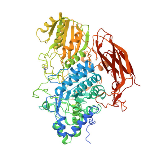
Structures of beta-glycosidase LXYL-P1-2 reveals the product binding state of GH3 family and a specific pocket for Taxol recognition.

Yang, L.Y.Chen, T.J.Wang, F.Li, L.Yu, W.B.Si, Y.K.Chen, J.J.Liu, W.C.Zhu, P.Gong, W.M.

(2020) Commun Biol 3: 22-22

  • DOI: https://doi.org/10.1038/s42003-019-0744-4
  • Primary Citation Related Structures: 
    6JBS, 6KJ0

  • PubMed Abstract: 

    LXYL-P1-2 is one of the few xylosidases that efficiently catalyze the reaction from 7-β-xylosyl-10-deacetyltaxol (XDT) to 10-deacetyltaxol (DT), and is a potential enzyme used in Taxol industrial production. Here we report the crystal structure of LXYL-P1-2 and its XDT binding complex. These structures reveal an enzyme/product complex with the sugar conformation different from the enzyme/substrate complex reported previously in GH3 enzymes, even in the whole glycohydrolases family. In addition, the DT binding pocket is identified as the structural basis for the substrate specificity. Further structure analysis reveals common features in LXYL-P1-2 and Taxol binding protein tubulin, which might provide useful information for designing new Taxol carrier proteins for drug delivery.


  • Organizational Affiliation
    • National Research Laboratory for Physical Sciences in Microscales, University of Science and Technology of China, 230026, Hefei, Anhui, China.

Macromolecule Content 

  • Total Structure Weight: 362.42 kDa 
  • Atom Count: 25,512 
  • Modeled Residue Count: 3,045 
  • Deposited Residue Count: 3,236 
  • Unique protein chains: 1

Macromolecules

Find similar proteins by:|  3D Structure
Entity ID: 1
MoleculeChains  Sequence LengthOrganismDetailsImage
Beta-D-xylosidase/beta-D-glucosidase
A, B, C, D
809Lentinula edodesMutation(s): 0 
Gene Names: Lxyl-p1-2
EC: 3.2.1.21
UniProt
Find proteins for G8GLP2 (Lentinula edodes)
Explore G8GLP2 
Go to UniProtKB:  G8GLP2
Entity Groups
Sequence Clusters30% Identity50% Identity70% Identity90% Identity95% Identity100% Identity
UniProt GroupG8GLP2
Glycosylation
Glycosylation Sites: 7
Sequence Annotations
Expand
Reference Sequence

Oligosaccharides

Help  
Entity ID: 2
MoleculeChains Length2D Diagram GlycosylationD Interactions
beta-D-mannopyranose-(1-4)-2-acetamido-2-deoxy-beta-D-glucopyranose-(1-4)-2-acetamido-2-deoxy-beta-D-glucopyranose
E, N
3N-Glycosylation
Glycosylation Resources
GlyTouCan: G15407YE
GlyCosmos: G15407YE
GlyGen: G15407YE
Entity ID: 3
MoleculeChains Length2D Diagram GlycosylationD Interactions
alpha-D-mannopyranose-(1-2)-beta-D-mannopyranose-(1-3)-[alpha-D-mannopyranose-(1-3)-alpha-D-mannopyranose-(1-6)]beta-D-mannopyranose-(1-4)-2-acetamido-2-deoxy-beta-D-glucopyranose-(1-4)-2-acetamido-2-deoxy-beta-D-glucopyranose
F
7N-Glycosylation
Glycosylation Resources
GlyTouCan: G90815BM
GlyCosmos: G90815BM
GlyGen: G90815BM
Entity ID: 4
MoleculeChains Length2D Diagram GlycosylationD Interactions
alpha-D-mannopyranose-(1-2)-alpha-D-mannopyranose-(1-2)-alpha-D-mannopyranose-(1-3)-alpha-D-mannopyranose-(1-4)-2-acetamido-2-deoxy-beta-D-glucopyranose-(1-4)-2-acetamido-2-deoxy-beta-D-glucopyranose
G
6N-Glycosylation
Glycosylation Resources
GlyTouCan: G60005GQ
GlyCosmos: G60005GQ
GlyGen: G60005GQ
Entity ID: 5
MoleculeChains Length2D Diagram GlycosylationD Interactions
alpha-D-mannopyranose-(1-4)-2-acetamido-2-deoxy-beta-D-glucopyranose-(1-4)-2-acetamido-2-deoxy-beta-D-glucopyranose
H, K
3N-Glycosylation
Glycosylation Resources
GlyTouCan: G62182OO
GlyCosmos: G62182OO
GlyGen: G62182OO
Entity ID: 6
MoleculeChains Length2D Diagram GlycosylationD Interactions
alpha-D-mannopyranose-(1-2)-alpha-D-mannopyranose-(1-3)-[alpha-D-mannopyranose-(1-3)-alpha-D-mannopyranose-(1-6)]beta-D-mannopyranose-(1-4)-2-acetamido-2-deoxy-beta-D-glucopyranose-(1-4)-2-acetamido-2-deoxy-beta-D-glucopyranose
I, L, O
7N-Glycosylation
Glycosylation Resources
GlyTouCan: G07617FP
GlyCosmos: G07617FP
GlyGen: G07617FP
Entity ID: 7
MoleculeChains Length2D Diagram GlycosylationD Interactions
2-acetamido-2-deoxy-beta-D-glucopyranose-(1-4)-2-acetamido-2-deoxy-beta-D-glucopyranose
J
2N-Glycosylation
Glycosylation Resources
GlyTouCan: G42666HT
GlyCosmos: G42666HT
GlyGen: G42666HT
Entity ID: 8
MoleculeChains Length2D Diagram GlycosylationD Interactions
beta-D-mannopyranose-(1-3)-[alpha-D-mannopyranose-(1-6)]alpha-D-mannopyranose-(1-4)-2-acetamido-2-deoxy-beta-D-glucopyranose-(1-4)-2-acetamido-2-deoxy-beta-D-glucopyranose
M
5N-Glycosylation
Glycosylation Resources
GlyTouCan: G66753FB
GlyCosmos: G66753FB
GlyGen: G66753FB
Entity ID: 9
MoleculeChains Length2D Diagram GlycosylationD Interactions
alpha-D-mannopyranose-(1-2)-alpha-D-mannopyranose-(1-2)-[alpha-D-mannopyranose-(1-6)]alpha-D-mannopyranose-(1-3)-alpha-D-mannopyranose-(1-4)-2-acetamido-2-deoxy-beta-D-glucopyranose-(1-4)-2-acetamido-2-deoxy-beta-D-glucopyranose
P
7N-Glycosylation
Glycosylation Resources
GlyTouCan: G40343UA
GlyCosmos: G40343UA
GlyGen: G40343UA

Small Molecules

Ligands 2 Unique
IDChains Name / Formula / InChI Key2D Diagram3D Interactions
NAG

Query on NAG


Download:Ideal Coordinates CCD File
AA [auth C]
BA [auth C]
CA [auth C]
DA [auth C]
FA [auth D]
AA [auth C],
BA [auth C],
CA [auth C],
DA [auth C],
FA [auth D],
GA [auth D],
HA [auth D],
IA [auth D],
Q [auth A],
R [auth A],
S [auth A],
T [auth A],
V [auth B],
W [auth B],
X [auth B],
Y [auth B]
2-acetamido-2-deoxy-beta-D-glucopyranose
C8 H15 N O6
OVRNDRQMDRJTHS-FMDGEEDCSA-N
TRS

Query on TRS


Download:Ideal Coordinates CCD File
EA [auth C],
JA [auth D],
U [auth A],
Z [auth B]
2-AMINO-2-HYDROXYMETHYL-PROPANE-1,3-DIOL
C4 H12 N O3
LENZDBCJOHFCAS-UHFFFAOYSA-O

Experimental Data & Validation

Experimental Data

  • Method: X-RAY DIFFRACTION
  • Resolution: 2.40 Å
  • R-Value Free:  0.251 (Depositor), 0.253 (DCC) 
  • R-Value Work:  0.184 (Depositor), 0.190 (DCC) 
  • R-Value Observed: 0.188 (Depositor) 
Space Group: P 43 21 2
Unit Cell:
Length ( Å )Angle ( ˚ )
a = 131.913α = 90
b = 131.913β = 90
c = 385.937γ = 90
Software Package:
Software NamePurpose
REFMACrefinement
HKL-2000data collection
HKL-2000data scaling
PHASERphasing

Structure Validation

View Full Validation Report


Ligand Structure Quality Assessment 


Entry History 

& Funding Information

Deposition Data


Funding OrganizationLocationGrant Number
National Natural Science Foundation of ChinaChina31270796

Revision History  (Full details and data files)

  • Version 1.0: 2020-02-12
    Type: Initial release
  • Version 1.1: 2020-02-26
    Changes: Database references
  • Version 2.0: 2020-07-29
    Type: Remediation
    Reason: Carbohydrate remediation
    Changes: Advisory, Atomic model, Data collection, Derived calculations, Structure summary
  • Version 2.1: 2024-11-13
    Changes: Data collection, Database references, Derived calculations, Structure summary