


Crystal structure of the flagellin protein FlaG from Helicobacter pylori.
Tsai, J.Y., Yeh, Y.H., Lin, L.D., Sun, Y.J., Hsiao, C.D.(2019) J Chin Chem Soc 66: 1177-1184
Experimental Data Snapshot
wwPDB Validation 3D Report Full Report

(2019) J Chin Chem Soc 66: 1177-1184
Entity ID: 1 | |||||
|---|---|---|---|---|---|
| Molecule | Chains | Sequence Length | Organism | Details | Image |
| Flagellar biosynthesis protein FlaG | 73 | Helicobacter pylori | Mutation(s): 0 Gene Names: BB430_02730, HPY1198_01455 | 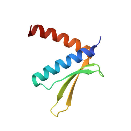 | |
| Length ( Å ) | Angle ( ˚ ) |
|---|---|
| a = 58.411 | α = 90 |
| b = 58.411 | β = 90 |
| c = 231.921 | γ = 120 |
| Software Name | Purpose |
|---|---|
| SCALEPACK | data scaling |
| PHENIX | refinement |
| PDB_EXTRACT | data extraction |
| Funding Organization | Location | Grant Number |
|---|---|---|
| Ministry of Science and Technology (Taiwan) | Taiwan | -- |


