6JU9 | pdb_00006ju9

Aspergillus oryzae active-tyrosinase copper-bound C92A mutant complexed with L-tyrosine


Experimental Data Snapshot

  • Method: X-RAY DIFFRACTION
  • Resolution: 1.42 Å
  • R-Value Free: 
    0.197 (Depositor), 0.206 (DCC) 
  • R-Value Work: 
    0.154 (Depositor) 
  • R-Value Observed: 
    0.156 (Depositor) 

Starting Model: experimental
View more details

wwPDB Validation 3D Report Full Report

Validation slider image for 6JU9

Ligand Structure Quality Assessment 


This is version 2.0 of the entry. See complete history

Literature

Copper-Oxygen Dynamics in the Tyrosinase Mechanism.

Fujieda, N.Umakoshi, K.Ochi, Y.Nishikawa, Y.Yanagisawa, S.Kubo, M.Kurisu, G.Itoh, S.

(2020) Angew Chem Int Ed Engl 59: 13385-13390

  • DOI: https://doi.org/10.1002/anie.202004733
  • Primary Citation Related Structures: 
    6JU4, 6JU5, 6JU6, 6JU7, 6JU8, 6JU9, 6JUA, 6JUB, 6JUC, 6JUD

  • PubMed Abstract: 

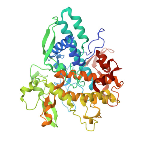
    The dinuclear copper enzyme, tyrosinase, activates O 2 to form a (μ-η 2 2 -peroxido)dicopper(II) species, which hydroxylates phenols to catechols. However, the exact mechanism of phenolase reaction in the catalytic site of tyrosinase is still under debate. We herein report the near atomic resolution X-ray crystal structures of the active tyrosinases with substrate l-tyrosine. At their catalytic sites, CuA moved toward l-tyrosine (CuA1 → CuA2), whose phenol oxygen directly coordinates to CuA2, involving the movement of CuB (CuB1 → CuB2). The crystal structures and spectroscopic analyses of the dioxygen-bound tyrosinases demonstrated that the peroxide ligand rotated, spontaneously weakening its O-O bond. Thus, the copper migration induced by the substrate-binding is accompanied by rearrangement of the bound peroxide species so as to provide one of the peroxide oxygen atoms with access to the phenol substrate's ϵ carbon atom.


  • Organizational Affiliation
    • Department of Applied Life Sciences, Graduate School of Life and Environmental Sciences, Osaka Prefecture University, 1-1 Gakuen-cho, Naka-ku, Sakai-shi, Osaka, 599-8531, Japan.

Macromolecule Content 

  • Total Structure Weight: 107.87 kDa 
  • Atom Count: 7,787 
  • Modeled Residue Count: 869 
  • Deposited Residue Count: 922 
  • Unique protein chains: 1

Macromolecules

Find similar proteins by:|  3D Structure
Entity ID: 1
MoleculeChains  Sequence LengthOrganismDetailsImage
Tyrosinase
A, B
461Aspergillus oryzaeMutation(s): 1 
Gene Names: OAory_01107480
EC: 1.14.18.1
UniProt
Find proteins for A0A1S9DK56 (Aspergillus oryzae)
Explore A0A1S9DK56 
Go to UniProtKB:  A0A1S9DK56
Entity Groups
Sequence Clusters30% Identity50% Identity70% Identity90% Identity95% Identity100% Identity
UniProt GroupA0A1S9DK56
Sequence Annotations
Expand
Reference Sequence

Small Molecules

Ligands 4 Unique
IDChains Name / Formula / InChI Key2D Diagram3D Interactions
DAH

Query on DAH


Download:Ideal Coordinates CCD File
D [auth A]3,4-DIHYDROXYPHENYLALANINE
C9 H11 N O4
WTDRDQBEARUVNC-LURJTMIESA-N
TYR

Query on TYR


Download:Ideal Coordinates CCD File
C [auth A],
H [auth B]
TYROSINE
C9 H11 N O3
OUYCCCASQSFEME-QMMMGPOBSA-N
CU

Query on CU


Download:Ideal Coordinates CCD File
E [auth A],
F [auth A],
I [auth B],
J [auth B]
COPPER (II) ION
Cu
JPVYNHNXODAKFH-UHFFFAOYSA-N
NO3

Query on NO3


Download:Ideal Coordinates CCD File
G [auth A]NITRATE ION
N O3
NHNBFGGVMKEFGY-UHFFFAOYSA-N

Experimental Data & Validation

Experimental Data

  • Method: X-RAY DIFFRACTION
  • Resolution: 1.42 Å
  • R-Value Free:  0.197 (Depositor), 0.206 (DCC) 
  • R-Value Work:  0.154 (Depositor) 
  • R-Value Observed: 0.156 (Depositor) 
Space Group: P 21 21 21
Unit Cell:
Length ( Å )Angle ( ˚ )
a = 54.544α = 90
b = 104.97β = 90
c = 154.877γ = 90
Software Package:
Software NamePurpose
DENZOdata reduction
HKL-2000data scaling
PHASERphasing
SHELXLrefinement

Structure Validation

View Full Validation Report


Ligand Structure Quality Assessment 


Entry History 

& Funding Information

Deposition Data


Funding OrganizationLocationGrant Number
Japan Science and TechnologyJapanCREST(JPMJCR16P1)

Revision History  (Full details and data files)

  • Version 1.0: 2020-05-13
    Type: Initial release
  • Version 1.1: 2020-08-12
    Changes: Database references, Derived calculations
  • Version 1.2: 2023-11-22
    Changes: Data collection, Database references, Refinement description
  • Version 2.0: 2025-09-17
    Changes: Atomic model, Data collection, Derived calculations, Structure summary