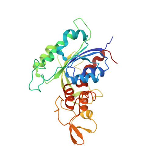
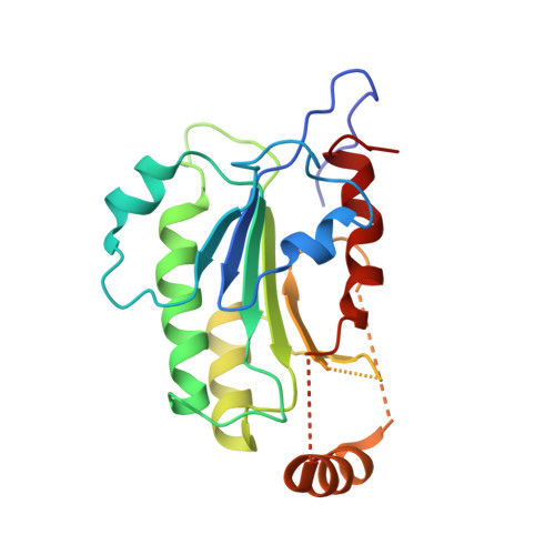
6KDL

Crystal structure of human DNMT3B-DNMT3L complex (I)


Experimental Data Snapshot

  • Method: X-RAY DIFFRACTION
  • Resolution: 3.27 Å
  • R-Value Free: 0.212 
  • R-Value Work: 0.179 
  • R-Value Observed: 0.180 

Starting Model: experimental
View more details

wwPDB Validation   3D Report Full Report


Ligand Structure Quality Assessment 


This is version 1.3 of the entry. See complete history


Literature

Structural insights into CpG-specific DNA methylation by human DNA methyltransferase 3B.

Lin, C.C.Chen, Y.P.Yang, W.Z.Shen, J.C.K.Yuan, H.S.

(2020) Nucleic Acids Res 48: 3949-3961

  • DOI: https://doi.org/10.1093/nar/gkaa111
  • Primary Citation of Related Structures:  
    6KDA, 6KDB, 6KDL, 6KDP, 6KDT

  • PubMed Abstract: 

    DNA methyltransferases are primary enzymes for cytosine methylation at CpG sites of epigenetic gene regulation in mammals. De novo methyltransferases DNMT3A and DNMT3B create DNA methylation patterns during development, but how they differentially implement genomic DNA methylation patterns is poorly understood. Here, we report crystal structures of the catalytic domain of human DNMT3B-3L complex, noncovalently bound with and without DNA of different sequences. Human DNMT3B uses two flexible loops to enclose DNA and employs its catalytic loop to flip out the cytosine base. As opposed to DNMT3A, DNMT3B specifically recognizes DNA with CpGpG sites via residues Asn779 and Lys777 in its more stable and well-ordered target recognition domain loop to facilitate processive methylation of tandemly repeated CpG sites. We also identify a proton wire water channel for the final deprotonation step, revealing the complete working mechanism for cytosine methylation by DNMT3B and providing the structural basis for DNMT3B mutation-induced hypomethylation in immunodeficiency, centromere instability and facial anomalies syndrome.


  • Organizational Affiliation

    Institute of Molecular Biology, Academia Sinica, Taipei 11529, Taiwan.


Macromolecules
Find similar proteins by:  (by identity cutoff)  |  3D Structure
Entity ID: 1
MoleculeChains Sequence LengthOrganismDetailsImage
DNA (cytosine-5)-methyltransferase 3B
A, D
286Homo sapiensMutation(s): 0 
Gene Names: DNMT3B
EC: 2.1.1.37
UniProt & NIH Common Fund Data Resources
Find proteins for Q9UBC3 (Homo sapiens)
Go to UniProtKB:  Q9UBC3
PHAROS:  Q9UBC3
GTEx:  ENSG00000088305 
Sequence Annotations
Expand
  • Reference Sequence
Find similar proteins by:  (by identity cutoff)  |  3D Structure
Entity ID: 2
MoleculeChains Sequence LengthOrganismDetailsImage
DNA (cytosine-5)-methyltransferase 3-like
B, C
204Homo sapiensMutation(s): 0 
Gene Names: DNMT3L
UniProt & NIH Common Fund Data Resources
Find proteins for Q9UJW3 (Homo sapiens)
Go to UniProtKB:  Q9UJW3
PHAROS:  Q9UJW3
GTEx:  ENSG00000142182 
Sequence Annotations
Expand
  • Reference Sequence
Small Molecules
Ligands 1 Unique
IDChains Name / Formula / InChI Key2D Diagram3D Interactions
SAH
Query on SAH

Download Ideal Coordinates CCD File 
E [auth A],
F [auth D]
S-ADENOSYL-L-HOMOCYSTEINE
C14 H20 N6 O5 S
ZJUKTBDSGOFHSH-WFMPWKQPSA-N
Binding Affinity Annotations 
IDSourceBinding Affinity
SAH BindingDB:  6KDL IC50: min: 200, max: 5.00e+4 (nM) from 4 assay(s)
Experimental Data & Validation

Experimental Data

  • Method: X-RAY DIFFRACTION
  • Resolution: 3.27 Å
  • R-Value Free: 0.212 
  • R-Value Work: 0.179 
  • R-Value Observed: 0.180 
  • Space Group: P 31
Unit Cell:
Length ( Å )Angle ( ˚ )
a = 192.932α = 90
b = 192.932β = 90
c = 50.041γ = 120
Software Package:
Software NamePurpose
PHENIXrefinement
HKL-2000data reduction
HKL-2000data scaling
PHASERphasing

Structure Validation

View Full Validation Report


Ligand Structure Quality Assessment 


Entry History 

Deposition Data

Revision History  (Full details and data files)

  • Version 1.0: 2020-02-19
    Type: Initial release
  • Version 1.1: 2020-03-04
    Changes: Database references
  • Version 1.2: 2020-04-22
    Changes: Database references
  • Version 1.3: 2023-11-22
    Changes: Data collection, Database references, Refinement description