6MON | pdb_00006mon

Crystal structure of human SMYD2 in complex with Nle-peptide inhibitor


Experimental Data Snapshot

  • Method: X-RAY DIFFRACTION
  • Resolution: 2.71 Å
  • R-Value Free: 
    0.227 (Depositor), 0.228 (DCC) 
  • R-Value Work: 
    0.210 (Depositor), 0.215 (DCC) 
  • R-Value Observed: 
    0.211 (Depositor) 

Starting Model: experimental
View more details

wwPDB Validation 3D Report Full Report

Validation slider image for 6MON

Ligand Structure Quality Assessment 


This is version 1.3 of the entry. See complete history

Literature

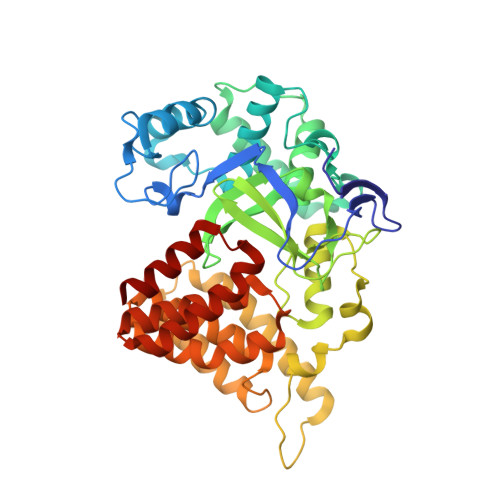
A functional proteomics platform to reveal the sequence determinants of lysine methyltransferase substrate selectivity.

Cornett, E.M.Dickson, B.M.Krajewski, K.Spellmon, N.Umstead, A.Vaughan, R.M.Shaw, K.M.Versluis, P.P.Cowles, M.W.Brunzelle, J.Yang, Z.Vega, I.E.Sun, Z.W.Rothbart, S.B.

(2018) Sci Adv 4: eaav2623-eaav2623

  • DOI: https://doi.org/10.1126/sciadv.aav2623
  • Primary Citation Related Structures: 
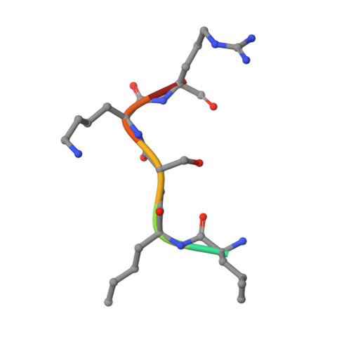
    6MON

  • PubMed Abstract: 

    Lysine methylation is a key regulator of histone protein function. Beyond histones, few connections have been made to the enzymes responsible for the deposition of these posttranslational modifications. Here, we debut a high-throughput functional proteomics platform that maps the sequence determinants of lysine methyltransferase (KMT) substrate selectivity without a priori knowledge of a substrate or target proteome. We demonstrate the predictive power of this approach for identifying KMT substrates, generating scaffolds for inhibitor design, and predicting the impact of missense mutations on lysine methylation signaling. By comparing KMT selectivity profiles to available lysine methylome datasets, we reveal a disconnect between preferred KMT substrates and the ability to detect these motifs using standard mass spectrometry pipelines. Collectively, our studies validate the use of this platform for guiding the study of lysine methylation signaling and suggest that substantial gaps exist in proteome-wide curation of lysine methylomes.


  • Organizational Affiliation
    • Center for Epigenetics, Van Andel Research Institute, Grand Rapids, MI 49503, USA.

Macromolecule Content 

  • Total Structure Weight: 101.55 kDa 
  • Atom Count: 7,074 
  • Modeled Residue Count: 870 
  • Deposited Residue Count: 872 
  • Unique protein chains: 2

Macromolecules

Find similar proteins by:|  3D Structure
Entity ID: 1
MoleculeChains  Sequence LengthOrganismDetailsImage
N-lysine methyltransferase SMYD2
A, B
429Homo sapiensMutation(s): 1 
Gene Names: SMYD2KMT3C
EC: 2.1.1 (PDB Primary Data), 2.1.1.43 (PDB Primary Data), 2.1.1.354 (UniProt)
UniProt & NIH Common Fund Data Resources
Find proteins for Q9NRG4 (Homo sapiens)
Explore Q9NRG4 
Go to UniProtKB:  Q9NRG4
PHAROS:  Q9NRG4
GTEx:  ENSG00000143499 
Entity Groups
Sequence Clusters30% Identity50% Identity70% Identity90% Identity95% Identity100% Identity
UniProt GroupQ9NRG4
Sequence Annotations
Expand
Reference Sequence
Find similar proteins by:  Sequence   |   3D Structure  
Entity ID: 2
MoleculeChains  Sequence LengthOrganismDetailsImage
LYS-LEU-NLE-SER-LYS-ARG-GLY
C, D
7synthetic constructMutation(s): 0 

Small Molecules

Ligands 3 Unique
IDChains Name / Formula / InChI Key2D Diagram3D Interactions
SAH

Query on SAH


Download:Ideal Coordinates CCD File
H [auth A],
M [auth B]
S-ADENOSYL-L-HOMOCYSTEINE
C14 H20 N6 O5 S
ZJUKTBDSGOFHSH-WFMPWKQPSA-N
GOL

Query on GOL


Download:Ideal Coordinates CCD File
I [auth A]GLYCEROL
C3 H8 O3
PEDCQBHIVMGVHV-UHFFFAOYSA-N
ZN

Query on ZN


Download:Ideal Coordinates CCD File
E [auth A]
F [auth A]
G [auth A]
J [auth B]
K [auth B]
E [auth A],
F [auth A],
G [auth A],
J [auth B],
K [auth B],
L [auth B]
ZINC ION
Zn
PTFCDOFLOPIGGS-UHFFFAOYSA-N
Modified Residues  1 Unique
IDChains TypeFormula2D DiagramParent
NLE
Query on NLE
C, D
L-PEPTIDE LINKINGC6 H13 N O2LEU

Experimental Data & Validation

Experimental Data

  • Method: X-RAY DIFFRACTION
  • Resolution: 2.71 Å
  • R-Value Free:  0.227 (Depositor), 0.228 (DCC) 
  • R-Value Work:  0.210 (Depositor), 0.215 (DCC) 
  • R-Value Observed: 0.211 (Depositor) 
Space Group: P 42
Unit Cell:
Length ( Å )Angle ( ˚ )
a = 153.728α = 90
b = 153.728β = 90
c = 53.457γ = 90
Software Package:
Software NamePurpose
PHASERphasing
PHENIXrefinement
PDB_EXTRACTdata extraction
XDSdata reduction
Aimlessdata scaling

Structure Validation

View Full Validation Report


Ligand Structure Quality Assessment 


Entry History 

Deposition Data

Revision History  (Full details and data files)

  • Version 1.0: 2018-12-12
    Type: Initial release
  • Version 1.1: 2023-10-11
    Changes: Data collection, Database references, Derived calculations, Refinement description
  • Version 1.2: 2023-11-15
    Changes: Data collection
  • Version 1.3: 2024-11-13
    Changes: Data collection, Structure summary