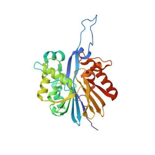
6N9Q

Structure of the Quorum Quenching lactonase from Parageobacillus caldoxylosilyticus bind to substrate C4-AHL


Experimental Data Snapshot

  • Method: X-RAY DIFFRACTION
  • Resolution: 2.35 Å
  • R-Value Free: 0.211 
  • R-Value Work: 0.166 
  • R-Value Observed: 0.168 

Starting Model: experimental
View more details

wwPDB Validation   3D Report Full Report


Ligand Structure Quality Assessment 


This is version 1.2 of the entry. See complete history


Literature

The Structural Determinants Accounting for the Broad Substrate Specificity of the Quorum Quenching Lactonase GcL.

Bergonzi, C.Schwab, M.Naik, T.Elias, M.

(2019) Chembiochem 20: 1848-1855

  • DOI: https://doi.org/10.1002/cbic.201900024
  • Primary Citation of Related Structures:  
    6N9I, 6N9Q, 6N9R

  • PubMed Abstract: 

    Quorum quenching lactonases are enzymes capable of hydrolyzing lactones, including N-acyl homoserine lactones (AHLs). AHLs are molecules known as signals in bacterial communication dubbed quorum sensing. Bacterial signal disruption by lactonases was previously reported to inhibit behavior regulated by quorum sensing, such as the expression of virulence factors and the formation of biofilms. Herein, we report the enzymatic and structural characterization of a novel lactonase representative from the metallo-β-lactamase superfamily, dubbed GcL. GcL is a broad spectrum and highly proficient lactonase, with k cat /K M values in the range of 10 4 to 10 6  m -1  s -1 . Analysis of free GcL structures and in complex with AHL substrates of different acyl chain length, namely, C4-AHL and 3-oxo-C12-AHL, allowed their respective binding modes to be elucidated. Structures reveal three subsites in the binding crevice: 1) the small subsite where chemistry is performed on the lactone ring; 2) a hydrophobic ring that accommodates the amide group of AHLs and small acyl chains; and 3) the outer, hydrophilic subsite that extends to the protein surface. Unexpectedly, the absence of structural accommodation for long substrate acyl chains seems to relate to the broad substrate specificity of the enzyme.


  • Organizational Affiliation

    Biochemistry, Molecular Biology and Biophysics Department and, BioTechnology Institute, University of Minnesota, Saint Paul, MN, 55108, USA.


Macromolecules
Find similar proteins by:  (by identity cutoff)  |  3D Structure
Entity ID: 1
MoleculeChains Sequence LengthOrganismDetailsImage
Lactonase GcLA [auth P],
B [auth D]
282Parageobacillus caldoxylosilyticus NBRC 107762Mutation(s): 0 
Gene Names: GCA01S_030_00190
UniProt
Find proteins for A0A023DFE8 (Parageobacillus caldoxylosilyticus NBRC 107762)
Explore A0A023DFE8 
Go to UniProtKB:  A0A023DFE8
Entity Groups  
Sequence Clusters30% Identity50% Identity70% Identity90% Identity95% Identity100% Identity
UniProt GroupA0A023DFE8
Sequence Annotations
Expand
  • Reference Sequence
Small Molecules
Ligands 8 Unique
IDChains Name / Formula / InChI Key2D Diagram3D Interactions
1PE
Query on 1PE

Download Ideal Coordinates CCD File 
GA [auth D],
HA [auth D],
N [auth P],
O [auth P]
PENTAETHYLENE GLYCOL
C10 H22 O6
JLFNLZLINWHATN-UHFFFAOYSA-N
HL4 (Subject of Investigation/LOI)
Query on HL4

Download Ideal Coordinates CCD File 
F [auth P],
X [auth D]
N-[(3S)-2-oxotetrahydrofuran-3-yl]butanamide
C8 H13 N O3
VFFNZZXXTGXBOG-LURJTMIESA-N
PGE
Query on PGE

Download Ideal Coordinates CCD File 
LA [auth D],
MA [auth D],
R [auth P],
S [auth P]
TRIETHYLENE GLYCOL
C6 H14 O4
ZIBGPFATKBEMQZ-UHFFFAOYSA-N
SO4
Query on SO4

Download Ideal Coordinates CCD File 
D [auth P],
E [auth P],
U [auth D],
V [auth D],
W [auth D]
SULFATE ION
O4 S
QAOWNCQODCNURD-UHFFFAOYSA-L
EDO
Query on EDO

Download Ideal Coordinates CCD File 
AA [auth D]
BA [auth D]
CA [auth D]
DA [auth D]
EA [auth D]
AA [auth D],
BA [auth D],
CA [auth D],
DA [auth D],
EA [auth D],
FA [auth D],
G [auth P],
H [auth P],
I [auth P],
J [auth P],
K [auth P],
L [auth P],
M [auth P],
Y [auth D],
Z [auth D]
1,2-ETHANEDIOL
C2 H6 O2
LYCAIKOWRPUZTN-UHFFFAOYSA-N
ACT
Query on ACT

Download Ideal Coordinates CCD File 
JA [auth D],
KA [auth D],
Q [auth P]
ACETATE ION
C2 H3 O2
QTBSBXVTEAMEQO-UHFFFAOYSA-M
CO
Query on CO

Download Ideal Coordinates CCD File 
C [auth P],
T [auth D]
COBALT (II) ION
Co
XLJKHNWPARRRJB-UHFFFAOYSA-N
FE
Query on FE

Download Ideal Coordinates CCD File 
IA [auth D],
P
FE (III) ION
Fe
VTLYFUHAOXGGBS-UHFFFAOYSA-N
Experimental Data & Validation

Experimental Data

  • Method: X-RAY DIFFRACTION
  • Resolution: 2.35 Å
  • R-Value Free: 0.211 
  • R-Value Work: 0.166 
  • R-Value Observed: 0.168 
  • Space Group: H 3
Unit Cell:
Length ( Å )Angle ( ˚ )
a = 108.02α = 90
b = 108.02β = 90
c = 222.14γ = 120
Software Package:
Software NamePurpose
REFMACrefinement
XDSdata reduction
XSCALEdata scaling
MOLREPphasing

Structure Validation

View Full Validation Report


Ligand Structure Quality Assessment 


Entry History 

Deposition Data

Revision History  (Full details and data files)

  • Version 1.0: 2019-04-03
    Type: Initial release
  • Version 1.1: 2019-08-14
    Changes: Data collection, Database references
  • Version 1.2: 2023-10-11
    Changes: Data collection, Database references, Derived calculations, Refinement description