6O8H | pdb_00006o8h

Crystal structure of UvrB mutant bound to duplex DNA


Experimental Data Snapshot

  • Method: X-RAY DIFFRACTION
  • Resolution: 2.39 Å
  • R-Value Free: 
    0.277 (Depositor), 0.277 (DCC) 
  • R-Value Work: 
    0.227 (Depositor), 0.227 (DCC) 
  • R-Value Observed: 
    0.230 (Depositor) 

wwPDB Validation 3D Report Full Report

Validation slider image for 6O8H

This is version 1.1 of the entry. See complete history

Literature

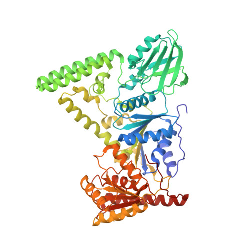
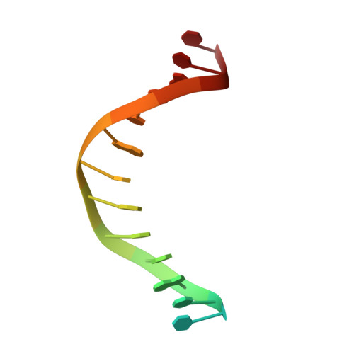
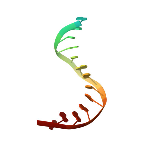
Mechanism of DNA Lesion Homing and Recognition by the Uvr Nucleotide Excision Repair System.

Lee, S.J.Sung, R.J.Verdine, G.L.

(2019) Research (Wash D C) 2019: 5641746-5641746

  • DOI: https://doi.org/10.34133/2019/5641746
  • Primary Citation Related Structures: 
    6O8E, 6O8F, 6O8G, 6O8H

  • PubMed Abstract: 

    Nucleotide excision repair (NER) is an essential DNA repair system distinguished from other such systems by its extraordinary versatility. NER removes a wide variety of structurally dissimilar lesions having only their bulkiness in common. NER can also repair several less bulky nucleobase lesions, such as 8-oxoguanine. Thus, how a single DNA repair system distinguishes such a diverse array of structurally divergent lesions from undamaged DNA has been one of the great unsolved mysteries in the field of genome maintenance. Here we employ a synthetic crystallography approach to obtain crystal structures of the pivotal NER enzyme UvrB in complex with duplex DNA, trapped at the stage of lesion-recognition. These structures coupled with biochemical studies suggest that UvrB integrates the ATPase-dependent helicase/translocase and lesion-recognition activities. Our work also conclusively establishes the identity of the lesion-containing strand and provides a compelling insight to how UvrB recognizes a diverse array of DNA lesions.


  • Organizational Affiliation
    • Department of Stem Cell and Regenerative Biology, Harvard University, Cambridge, MA 02138, USA.

Macromolecule Content 

  • Total Structure Weight: 78.14 kDa 
  • Atom Count: 5,323 
  • Modeled Residue Count: 597 
  • Deposited Residue Count: 625 
  • Unique protein chains: 1
  • Unique nucleic acid chains: 2

Macromolecules


Find similar proteins by:|  3D Structure
Entity ID: 1
MoleculeChains  Sequence LengthOrganismDetailsImage
UvrABC system protein B593[Bacillus] caldotenaxMutation(s): 0 
Gene Names: uvrB
UniProt
Find proteins for P56981 (Bacillus caldotenax)
Explore P56981 
Go to UniProtKB:  P56981
Entity Groups
Sequence Clusters30% Identity50% Identity70% Identity90% Identity95% Identity100% Identity
UniProt GroupP56981
Sequence Annotations
Expand
Reference Sequence
Find similar nucleic acids by:  Sequence
Entity ID: 2
MoleculeChains LengthOrganismImage
DNA (5'-D(P*CP*CP*AP*TP*CP*GP*CP*GP*CP*TP*AP*CP*C)-3')16synthetic construct
Sequence Annotations
Expand
Reference Sequence
Find similar nucleic acids by:  Sequence
Entity ID: 3
MoleculeChains LengthOrganismImage
DNA (5'-D(P*AP*GP*CP*GP*CP*GP*AP*TP*GP*GP*AP*GP*A)-3')16synthetic construct
Sequence Annotations
Expand
Reference Sequence

Experimental Data & Validation

Experimental Data

  • Method: X-RAY DIFFRACTION
  • Resolution: 2.39 Å
  • R-Value Free:  0.277 (Depositor), 0.277 (DCC) 
  • R-Value Work:  0.227 (Depositor), 0.227 (DCC) 
  • R-Value Observed: 0.230 (Depositor) 
Space Group: P 41 21 2
Unit Cell:
Length ( Å )Angle ( ˚ )
a = 124.791α = 90
b = 124.791β = 90
c = 96.175γ = 90
Software Package:
Software NamePurpose
REFMACrefinement
PDB_EXTRACTdata extraction
XDSdata reduction
SCALAdata scaling
PHASERphasing

Structure Validation

View Full Validation Report


Entry History 

& Funding Information

Deposition Data


Funding OrganizationLocationGrant Number
National Institutes of Health/National Cancer Institute (NIH/NCI)United States--

Revision History  (Full details and data files)

  • Version 1.0: 2020-01-22
    Type: Initial release
  • Version 1.1: 2024-03-13
    Changes: Advisory, Data collection, Database references