6ODD

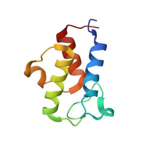
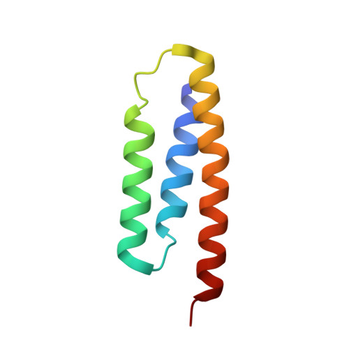
Crystal structure of the human complex ACP-ISD11


Experimental Data Snapshot

  • Method: X-RAY DIFFRACTION
  • Resolution: 2.00 Å
  • R-Value Free: 0.216 
  • R-Value Work: 0.193 
  • R-Value Observed: 0.194 

Starting Model: experimental
View more details

wwPDB Validation   3D Report Full Report


Ligand Structure Quality Assessment 


This is version 1.2 of the entry. See complete history


Literature

Structure of the Human ACP-ISD11 Heterodimer.

Herrera, M.G.Noguera, M.E.Sewell, K.E.Agudelo Suarez, W.A.Capece, L.Klinke, S.Santos, J.

(2019) Biochemistry 58: 4596-4609

  • DOI: https://doi.org/10.1021/acs.biochem.9b00539
  • Primary Citation of Related Structures:  
    6ODD

  • PubMed Abstract: 

    In recent years, the mammalian mitochondrial protein complex for iron-sulfur cluster assembly has been the focus of important studies. This is partly because of its high degree of relevance in cell metabolism and because mutations of the involved proteins are the cause of several human diseases. Cysteine desulfurase NFS1 is the key enzyme of the complex. At present, it is well-known that the active form of NFS1 is stabilized by the small protein ISD11. In this work, the structure of the human mitochondrial ACP-ISD11 heterodimer was determined at 2.0 Å resolution. ACP-ISD11 forms a cooperative unit stabilized by several ionic interactions, hydrogen bonds, and apolar interactions. The 4'-phosphopantetheine-acyl chain, which is covalently bound to ACP, interacts with several residues of ISD11, modulating together with ACP the foldability of ISD11. Recombinant human ACP-ISD11 was able to interact with the NFS1 desulfurase, thus yielding an active enzyme, and the NFS1/ACP-ISD11 core complex was activated by frataxin and ISCU proteins. Internal motions of ACP-ISD11 were studied by molecular dynamics simulations, showing the persistence of the interactions between both protein chains. The conformation of the dimer is similar to that found in the context of the (NFS1/ACP-ISD11) 2 supercomplex core, which contains the Escherichia coli ACP instead of the human variant. This fact suggests a sequential mechanism for supercomplex consolidation, in which the ACP-ISD11 complex may fold independently and, after that, the NFS1 dimer would be stabilized.


  • Organizational Affiliation

    Departamento de Fisiología y Biología Molecular y Celular, Facultad de Ciencias Exactas y Naturales , Universidad de Buenos Aires, Instituto de Biociencias, Biotecnología y Biomedicina (iB3), Intendente Güiraldes 2160-Ciudad Universitaria , C1428EGA Buenos Aires , Argentina.


Macromolecules
Find similar proteins by:  (by identity cutoff)  |  3D Structure
Entity ID: 1
MoleculeChains Sequence LengthOrganismDetailsImage
Acyl carrier protein, mitochondrial85Homo sapiensMutation(s): 0 
Gene Names: NDUFAB1
UniProt & NIH Common Fund Data Resources
Find proteins for O14561 (Homo sapiens)
Explore O14561 
Go to UniProtKB:  O14561
PHAROS:  O14561
GTEx:  ENSG00000004779 
Entity Groups  
Sequence Clusters30% Identity50% Identity70% Identity90% Identity95% Identity100% Identity
UniProt GroupO14561
Sequence Annotations
Expand
  • Reference Sequence
Find similar proteins by:  (by identity cutoff)  |  3D Structure
Entity ID: 2
MoleculeChains Sequence LengthOrganismDetailsImage
LYR motif-containing protein 474Homo sapiensMutation(s): 0 
Gene Names: LYRM4C6orf149ISD11CGI-203
UniProt & NIH Common Fund Data Resources
Find proteins for Q9HD34 (Homo sapiens)
Explore Q9HD34 
Go to UniProtKB:  Q9HD34
PHAROS:  Q9HD34
GTEx:  ENSG00000214113 
Entity Groups  
Sequence Clusters30% Identity50% Identity70% Identity90% Identity95% Identity100% Identity
UniProt GroupQ9HD34
Sequence Annotations
Expand
  • Reference Sequence
Small Molecules
Ligands 2 Unique
IDChains Name / Formula / InChI Key2D Diagram3D Interactions
8Q1
Query on 8Q1

Download Ideal Coordinates CCD File 
C [auth A]S-[2-({N-[(2R)-2-hydroxy-3,3-dimethyl-4-(phosphonooxy)butanoyl]-beta-alanyl}amino)ethyl] dodecanethioate
C23 H45 N2 O8 P S
MVHUOSAYFQKAMT-NRFANRHFSA-N
CA
Query on CA

Download Ideal Coordinates CCD File 
D [auth A]CALCIUM ION
Ca
BHPQYMZQTOCNFJ-UHFFFAOYSA-N
Experimental Data & Validation

Experimental Data

  • Method: X-RAY DIFFRACTION
  • Resolution: 2.00 Å
  • R-Value Free: 0.216 
  • R-Value Work: 0.193 
  • R-Value Observed: 0.194 
  • Space Group: I 21 3
Unit Cell:
Length ( Å )Angle ( ˚ )
a = 123.19α = 90
b = 123.19β = 90
c = 123.19γ = 90
Software Package:
Software NamePurpose
PHENIXrefinement
XDSdata reduction
XDSdata scaling
PDB_EXTRACTdata extraction
MoRDaphasing

Structure Validation

View Full Validation Report


Ligand Structure Quality Assessment 


Entry History & Funding Information

Deposition Data


Funding OrganizationLocationGrant Number
Friedreichs Ataxia Research Alliance (FARA)United States--

Revision History  (Full details and data files)

  • Version 1.0: 2019-11-27
    Type: Initial release
  • Version 1.1: 2019-12-04
    Changes: Database references
  • Version 1.2: 2023-10-11
    Changes: Data collection, Database references, Derived calculations, Refinement description