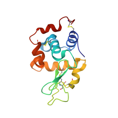
6PBB

Crystal structure of Hen Egg White Lysozyme in complex with I3C

  • Classification: HYDROLASE
  • Organism(s): Gallus gallus
  • Mutation(s): No 

  • Deposited: 2019-06-13 Released: 2019-07-10 
  • Deposition Author(s): Truong, J.Q.
  • Funding Organization(s): Australian Research Council (ARC)

Experimental Data Snapshot

  • Method: X-RAY DIFFRACTION
  • Resolution: 1.89 Å
  • R-Value Free: 0.240 
  • R-Value Work: 0.197 
  • R-Value Observed: 0.201 

wwPDB Validation   3D Report Full Report


Ligand Structure Quality Assessment 


This is version 1.4 of the entry. See complete history


Literature

Combining random microseed matrix screening and the magic triangle for the efficient structure solution of a potential lysin from bacteriophage P68.

Truong, J.Q.Panjikar, S.Shearwin-Whyatt, L.Bruning, J.B.Shearwin, K.E.

(2019) Acta Crystallogr D Struct Biol 75: 670-681

  • DOI: https://doi.org/10.1107/S2059798319009008
  • Primary Citation of Related Structures:  
    6O43, 6PBB

  • PubMed Abstract: 

    Two commonly encountered bottlenecks in the structure determination of a protein by X-ray crystallography are screening for conditions that give high-quality crystals and, in the case of novel structures, finding derivatization conditions for experimental phasing. In this study, the phasing molecule 5-amino-2,4,6-triiodoisophthalic acid (I3C) was added to a random microseed matrix screen to generate high-quality crystals derivatized with I3C in a single optimization experiment. I3C, often referred to as the magic triangle, contains an aromatic ring scaffold with three bound I atoms. This approach was applied to efficiently phase the structures of hen egg-white lysozyme and the N-terminal domain of the Orf11 protein from Staphylococcus phage P68 (Orf11 NTD) using SAD phasing. The structure of Orf11 NTD suggests that it may play a role as a virion-associated lysin or endolysin.


  • Organizational Affiliation

    School of Biological Sciences, The University of Adelaide, North Terrace, Adelaide, South Australia 5005, Australia.


Macromolecules
Find similar proteins by:  (by identity cutoff)  |  3D Structure
Entity ID: 1
MoleculeChains Sequence LengthOrganismDetailsImage
Lysozyme129Gallus gallusMutation(s): 0 
EC: 3.2.1.17
UniProt
Find proteins for P00698 (Gallus gallus)
Explore P00698 
Go to UniProtKB:  P00698
Entity Groups  
Sequence Clusters30% Identity50% Identity70% Identity90% Identity95% Identity100% Identity
UniProt GroupP00698
Sequence Annotations
Expand
  • Reference Sequence
Experimental Data & Validation

Experimental Data

  • Method: X-RAY DIFFRACTION
  • Resolution: 1.89 Å
  • R-Value Free: 0.240 
  • R-Value Work: 0.197 
  • R-Value Observed: 0.201 
  • Space Group: P 43 21 2
Unit Cell:
Length ( Å )Angle ( ˚ )
a = 77.16α = 90
b = 77.16β = 90
c = 38.201γ = 90
Software Package:
Software NamePurpose
PHENIXrefinement
XDSdata reduction
Aimlessdata scaling
Auto-Rickshawphasing

Structure Validation

View Full Validation Report


Ligand Structure Quality Assessment 


Entry History & Funding Information

Deposition Data

  • Released Date: 2019-07-10 
  • Deposition Author(s): Truong, J.Q.

Funding OrganizationLocationGrant Number
Australian Research Council (ARC)AustraliaDP150103009
Australian Research Council (ARC)AustraliaDP160101450

Revision History  (Full details and data files)

  • Version 1.0: 2019-07-10
    Type: Initial release
  • Version 1.1: 2019-07-17
    Changes: Data collection, Database references
  • Version 1.2: 2019-07-24
    Changes: Data collection, Database references
  • Version 1.3: 2020-01-01
    Changes: Author supporting evidence
  • Version 1.4: 2024-10-16
    Changes: Data collection, Database references, Structure summary