


Crystal structure of HEMK2-TRMT112 complex
Dong, C., Tempel, W., Bountra, C., Arrowsmith, C.H., Edwards, A.M., Min, J.To be published.
Experimental Data Snapshot
Starting Model: experimental
View more details
Entity ID: 1 | |||||
|---|---|---|---|---|---|
| Molecule | Chains | Sequence Length | Organism | Details | Image |
| Methyltransferase N6AMT1 | 233 | Homo sapiens | Mutation(s): 0 Gene Names: N6AMT1, C21orf127, HEMK2, PRED28 EC: 2.1.1 (PDB Primary Data), 2.1.1.72 (PDB Primary Data) | 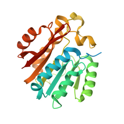 | |
UniProt & NIH Common Fund Data Resources | |||||
Find proteins for Q9Y5N5 (Homo sapiens) Explore Q9Y5N5 Go to UniProtKB: Q9Y5N5 | |||||
PHAROS: Q9Y5N5 GTEx: ENSG00000156239 | |||||
Entity Groups | |||||
| Sequence Clusters | 30% Identity50% Identity70% Identity90% Identity95% Identity100% Identity | ||||
| UniProt Group | Q9Y5N5 | ||||
Sequence AnnotationsExpand | |||||
| |||||
Entity ID: 2 | |||||
|---|---|---|---|---|---|
| Molecule | Chains | Sequence Length | Organism | Details | Image |
| Multifunctional methyltransferase subunit TRM112-like protein | 143 | Homo sapiens | Mutation(s): 0 Gene Names: TRMT112, AD-001, HSPC152, HSPC170 | 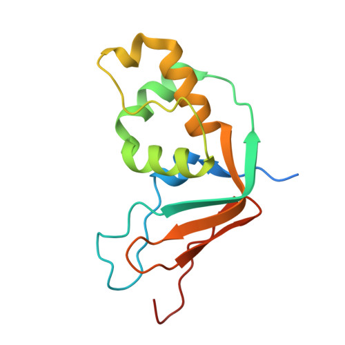 | |
UniProt & NIH Common Fund Data Resources | |||||
Find proteins for Q9UI30 (Homo sapiens) Explore Q9UI30 Go to UniProtKB: Q9UI30 | |||||
PHAROS: Q9UI30 GTEx: ENSG00000173113 | |||||
Entity Groups | |||||
| Sequence Clusters | 30% Identity50% Identity70% Identity90% Identity95% Identity100% Identity | ||||
| UniProt Group | Q9UI30 | ||||
Sequence AnnotationsExpand | |||||
| |||||
| Ligands 2 Unique | |||||
|---|---|---|---|---|---|
| ID | Chains | Name / Formula / InChI Key | 2D Diagram | 3D Interactions | |
| SAH Query on SAH | C [auth A] | S-ADENOSYL-L-HOMOCYSTEINE C14 H20 N6 O5 S ZJUKTBDSGOFHSH-WFMPWKQPSA-N |  | ||
| UNX Query on UNX | D [auth A] E [auth A] F [auth A] G [auth A] H [auth A] | UNKNOWN ATOM OR ION X |  | ||
| Length ( Å ) | Angle ( ˚ ) |
|---|---|
| a = 110.539 | α = 90 |
| b = 110.539 | β = 90 |
| c = 131.972 | γ = 120 |
| Software Name | Purpose |
|---|---|
| REFMAC | refinement |
| Aimless | data scaling |
| PDB_EXTRACT | data extraction |
| XDS | data reduction |
| REFMAC | phasing |


