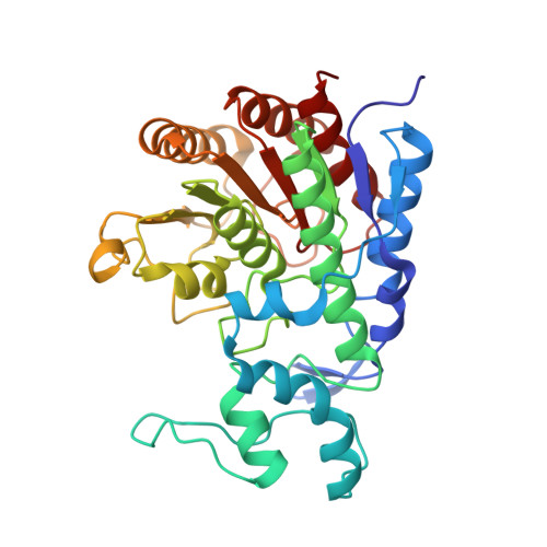
6PHZ

Crystal structure of Marinobacter subterrani acetylpolyamine amidohydrolase (msAPAH) complexed with 7-[(3-aminopropyl)amino]-1,1,1-trifluoroheptan-2-one


Experimental Data Snapshot

  • Method: X-RAY DIFFRACTION
  • Resolution: 2.00 Å
  • R-Value Free: 0.212 
  • R-Value Work: 0.161 
  • R-Value Observed: 0.164 

Starting Model: experimental
View more details

wwPDB Validation   3D Report Full Report


Ligand Structure Quality Assessment 


This is version 1.3 of the entry. See complete history


Literature

Structure and Function of the Acetylpolyamine Amidohydrolase from the Deep Earth HalophileMarinobacter subterrani.

Osko, J.D.Roose, B.W.Shinsky, S.A.Christianson, D.W.

(2019) Biochemistry 58: 3755-3766

  • DOI: https://doi.org/10.1021/acs.biochem.9b00582
  • Primary Citation of Related Structures:  
    6PHR, 6PHT, 6PHZ, 6PI1, 6PI8, 6PIA, 6PIC, 6PID

  • PubMed Abstract: 

    Polyamines are small organic cations that are essential for cellular function in all kingdoms of life. Polyamine metabolism is regulated by enzyme-catalyzed acetylation-deacetylation cycles in a fashion similar to the epigenetic regulation of histone function in eukaryotes. Bacterial polyamine deacetylases are particularly intriguing, because these enzymes share the fold and function of eukaryotic histone deacetylases. Recently, acetylpolyamine amidohydrolase from the deep earth halophile Marinobacter subterrani (msAPAH) was described. This Zn 2+ -dependent deacetylase shares 53% amino acid sequence identity with the acetylpolyamine amidohydrolase from Mycoplana ramosa (mrAPAH) and 22% amino acid sequence identity with the catalytic domain of histone deacetylase 10 from Danio rerio (zebrafish; zHDAC10), the eukaryotic polyamine deacetylase. The X-ray crystal structure of msAPAH, determined in complexes with seven different inhibitors as well as the acetate coproduct, shows how the chemical strategy of Zn 2+ -dependent amide hydrolysis and the catalytic specificity for cationic polyamine substrates is conserved in a subterranean halophile. Structural comparisons with mrAPAH reveal that an array of aspartate and glutamate residues unique to msAPAH enable the binding of one or more Mg 2+ ions in the active site and elsewhere on the protein surface. Notwithstanding these differences, activity assays with a panel of acetylpolyamine and acetyllysine substrates confirm that msAPAH is a broad-specificity polyamine deacetylase, much like mrAPAH. The broad substrate specificity contrasts with the narrow substrate specificity of zHDAC10, which is highly specific for N 8 -acetylspermidine hydrolysis. Notably, quaternary structural features govern the substrate specificity of msAPAH and mrAPAH, whereas tertiary structural features govern the substrate specificity of zHDAC10.


  • Organizational Affiliation

    Roy and Diana Vagelos Laboratories, Department of Chemistry , University of Pennsylvania , 231 South 34th Street , Philadelphia , Pennsylvania 19104-6323 , United States.


Macromolecules
Find similar proteins by:  (by identity cutoff)  |  3D Structure
Entity ID: 1
MoleculeChains Sequence LengthOrganismDetailsImage
Acetylpolyamine Amidohydrolase
A, B
348Marinobacter subterraniMutation(s): 0 
Gene Names: Msub_13096
UniProt
Find proteins for A0A0J7JFD7 (Marinobacter subterrani)
Explore A0A0J7JFD7 
Go to UniProtKB:  A0A0J7JFD7
Entity Groups  
Sequence Clusters30% Identity50% Identity70% Identity90% Identity95% Identity100% Identity
UniProt GroupA0A0J7JFD7
Sequence Annotations
Expand
  • Reference Sequence
Small Molecules
Ligands 4 Unique
IDChains Name / Formula / InChI Key2D Diagram3D Interactions
FKS (Subject of Investigation/LOI)
Query on FKS

Download Ideal Coordinates CCD File 
H [auth A],
N [auth B]
7-[(3-aminopropyl)amino]-1,1,1-trifluoroheptane-2,2-diol
C10 H21 F3 N2 O2
IVRAKANOVJJYCX-UHFFFAOYSA-N
ZN
Query on ZN

Download Ideal Coordinates CCD File 
E [auth A],
K [auth B]
ZINC ION
Zn
PTFCDOFLOPIGGS-UHFFFAOYSA-N
K
Query on K

Download Ideal Coordinates CCD File 
C [auth A],
D [auth A],
I [auth B],
J [auth B]
POTASSIUM ION
K
NPYPAHLBTDXSSS-UHFFFAOYSA-N
MG
Query on MG

Download Ideal Coordinates CCD File 
F [auth A],
G [auth A],
L [auth B],
M [auth B]
MAGNESIUM ION
Mg
JLVVSXFLKOJNIY-UHFFFAOYSA-N
Experimental Data & Validation

Experimental Data

  • Method: X-RAY DIFFRACTION
  • Resolution: 2.00 Å
  • R-Value Free: 0.212 
  • R-Value Work: 0.161 
  • R-Value Observed: 0.164 
  • Space Group: P 1 21 1
Unit Cell:
Length ( Å )Angle ( ˚ )
a = 47.37α = 90
b = 82.36β = 98.22
c = 90.72γ = 90
Software Package:
Software NamePurpose
PHENIXrefinement
MOSFLMdata reduction
Aimlessdata scaling
PHASERphasing

Structure Validation

View Full Validation Report


Ligand Structure Quality Assessment 


Entry History & Funding Information

Deposition Data


Funding OrganizationLocationGrant Number
National Institutes of Health/National Human Genome Research Institute (NIH/NHGRI)United StatesGM49758

Revision History  (Full details and data files)

  • Version 1.0: 2019-09-18
    Type: Initial release
  • Version 1.1: 2019-09-25
    Changes: Data collection, Database references
  • Version 1.2: 2019-12-18
    Changes: Author supporting evidence
  • Version 1.3: 2023-10-11
    Changes: Data collection, Database references, Derived calculations, Refinement description