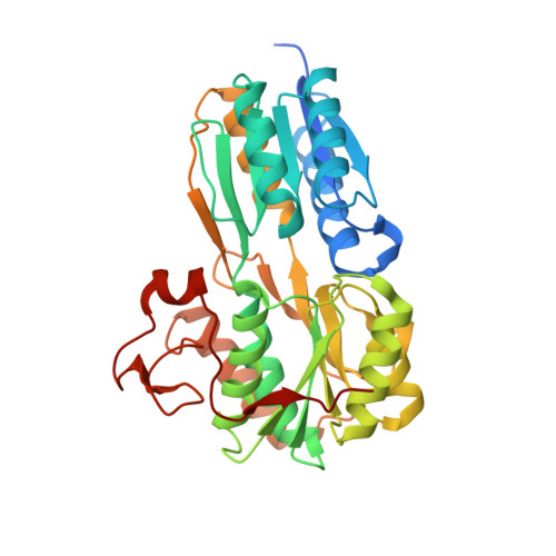
The evolving story of AtzT, a periplasmic binding protein.
Dennis, M.L., Esquirol, L., Nebl, T., Newman, J., Scott, C., Peat, T.S.(2019) Acta Crystallogr D Struct Biol 75: 995-1002
- PubMed: 31692473
- DOI: https://doi.org/10.1107/S2059798319013883
- Primary Citation of Related Structures:
6PI5, 6PI6, 6PII - PubMed Abstract:
Atrazine is an s-triazine-based herbicide that is used in many countries around the world in many millions of tons per year. A small number of organisms, such as Pseudomonas sp. strain ADP, have evolved to use this modified s-triazine as a food source, and the various genes required to metabolize atrazine can be found on a single plasmid. The atomic structures of seven of the eight proteins involved in the breakdown of atrazine by Pseudomonas sp. strain ADP have been determined by X-ray crystallography, but the structures of the proteins required by the cell to import atrazine for use as an energy source are still lacking. The structure of AtzT, a periplasmic binding protein that may be involved in the transport of a derivative of atrazine, 2-hydroxyatrazine, into the cell for mineralization, has now been determined. The structure was determined by SAD phasing using an ethylmercury phosphate derivative that diffracted X-rays to beyond 1.9 Å resolution. `Native' (guanine-bound) and 2-hydroxyatrazine-bound structures were also determined to high resolution (1.67 and 1.65 Å, respectively), showing that 2-hydroxyatrazine binds in a similar way to the purportedly native ligand. Structural similarities led to the belief that it may be possible to evolve AtzT from a purine-binding protein to a protein that can bind and detect atrazine in the environment.
Organizational Affiliation:
Biomedical Manufacturing Program, CSIRO, 343 Royal Parade, Parkville, VIC 3052, Australia.


