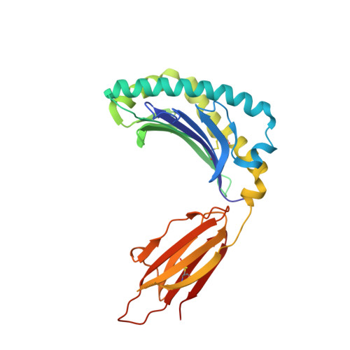
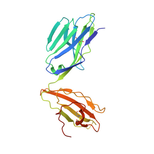
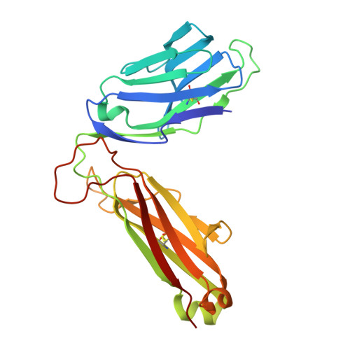
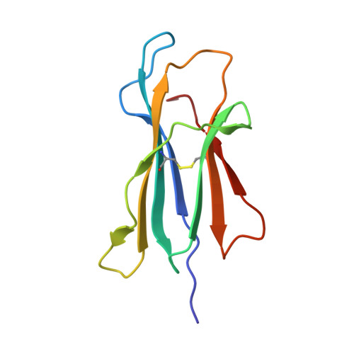
6PUJ

Structure of human MAIT A-F7 TCR in complex with human MR1-3`OH-Propyl-5-OP-U


Experimental Data Snapshot

  • Method: X-RAY DIFFRACTION
  • Resolution: 1.92 Å
  • R-Value Free: 0.194 
  • R-Value Work: 0.163 
  • R-Value Observed: 0.164 

Starting Model: experimental
View more details

wwPDB Validation   3D Report Full Report


Ligand Structure Quality Assessment 


This is version 1.5 of the entry. See complete history


Literature

The molecular basis underpinning the potency and specificity of MAIT cell antigens.

Awad, W.Ler, G.J.M.Xu, W.Keller, A.N.Mak, J.Y.W.Lim, X.Y.Liu, L.Eckle, S.B.G.Le Nours, J.McCluskey, J.Corbett, A.J.Fairlie, D.P.Rossjohn, J.

(2020) Nat Immunol 21: 400-411

  • DOI: https://doi.org/10.1038/s41590-020-0616-6
  • Primary Citation of Related Structures:  
    6PUC, 6PUD, 6PUE, 6PUF, 6PUG, 6PUH, 6PUI, 6PUJ, 6PUK, 6PUL, 6PUM

  • PubMed Abstract: 

    Mucosal-associated invariant T (MAIT) cells are activated by microbial riboflavin-based metabolite antigens when presented by MR1. How modifications to the potent antigen 5-OP-RU affect presentation by MR1 and MAIT cell activation remains unclear. Here we design 20 derivatives, termed altered metabolite ligands (AMLs), to dissect the impact of different antigen components on the human MAIT-MR1 axis. Analysis of 11 crystal structures of MAIT T cell antigen receptor (TCR)-MR1-AML ternary complexes, along with biochemical and functional assays, shows that MR1 cell-surface upregulation is influenced by ribityl and non-ribityl components of the ligand and the hydrophobicity of the MR1-AML interface. The polar ribityl chain of the AML strongly influences MAIT cell activation potency through dynamic compensatory interactions within a MAIT TCR-MR1-AML interaction triad. We define the basis by which the MAIT TCR can differentially recognize AMLs, thereby providing insight into MAIT cell antigen specificity and potency.


  • Organizational Affiliation

    Infection and Immunity Program and Department of Biochemistry and Molecular Biology, Biomedicine Discovery Institute, Monash University, Clayton, Victoria, Australia.


Macromolecules
Find similar proteins by:  (by identity cutoff)  |  3D Structure
Entity ID: 1
MoleculeChains Sequence LengthOrganismDetailsImage
Major histocompatibility complex class I-related gene protein
A, C
271Homo sapiensMutation(s): 0 
Gene Names: MR1
UniProt & NIH Common Fund Data Resources
Find proteins for Q95460 (Homo sapiens)
Explore Q95460 
Go to UniProtKB:  Q95460
PHAROS:  Q95460
GTEx:  ENSG00000153029 
Entity Groups  
Sequence Clusters30% Identity50% Identity70% Identity90% Identity95% Identity100% Identity
UniProt GroupQ95460
Sequence Annotations
Expand
  • Reference Sequence
Find similar proteins by:  (by identity cutoff)  |  3D Structure
Entity ID: 2
MoleculeChains Sequence LengthOrganismDetailsImage
Human TCR alpha chain
B, D
204Homo sapiensMutation(s): 0 
Gene Names: TRA@
Entity Groups  
Sequence Clusters30% Identity50% Identity70% Identity90% Identity95% Identity100% Identity
Sequence Annotations
Expand
  • Reference Sequence
Find similar proteins by:  (by identity cutoff)  |  3D Structure
Entity ID: 3
MoleculeChains Sequence LengthOrganismDetailsImage
Human TCR beta chain
E, G
246Homo sapiensMutation(s): 0 
Entity Groups  
Sequence Clusters30% Identity50% Identity70% Identity90% Identity95% Identity100% Identity
Sequence Annotations
Expand
  • Reference Sequence
Find similar proteins by:  (by identity cutoff)  |  3D Structure
Entity ID: 4
MoleculeChains Sequence LengthOrganismDetailsImage
Beta-2-microglobulin
F, H
100Homo sapiensMutation(s): 0 
Gene Names: B2MCDABP0092HDCMA22P
UniProt & NIH Common Fund Data Resources
Find proteins for P61769 (Homo sapiens)
Explore P61769 
Go to UniProtKB:  P61769
PHAROS:  P61769
GTEx:  ENSG00000166710 
Entity Groups  
Sequence Clusters30% Identity50% Identity70% Identity90% Identity95% Identity100% Identity
UniProt GroupP61769
Sequence Annotations
Expand
  • Reference Sequence
Small Molecules
Ligands 4 Unique
IDChains Name / Formula / InChI Key2D Diagram3D Interactions
Q7S (Subject of Investigation/LOI)
Query on Q7S

Download Ideal Coordinates CCD File 
K [auth A],
M [auth C]
6-[(3-hydroxypropyl)amino]-5-[(E)-(2-oxopropylidene)amino]pyrimidine-2,4(1H,3H)-dione
C10 H14 N4 O4
RCQDRIITDZKOID-LFYBBSHMSA-N
GOL
Query on GOL

Download Ideal Coordinates CCD File 
I [auth A]GLYCEROL
C3 H8 O3
PEDCQBHIVMGVHV-UHFFFAOYSA-N
CL
Query on CL

Download Ideal Coordinates CCD File 
J [auth A],
L [auth C]
CHLORIDE ION
Cl
VEXZGXHMUGYJMC-UHFFFAOYSA-M
NA
Query on NA

Download Ideal Coordinates CCD File 
N [auth E],
O [auth G]
SODIUM ION
Na
FKNQFGJONOIPTF-UHFFFAOYSA-N
Experimental Data & Validation

Experimental Data

  • Method: X-RAY DIFFRACTION
  • Resolution: 1.92 Å
  • R-Value Free: 0.194 
  • R-Value Work: 0.163 
  • R-Value Observed: 0.164 
  • Space Group: C 1 2 1
Unit Cell:
Length ( Å )Angle ( ˚ )
a = 218.607α = 90
b = 70.776β = 104.562
c = 143.802γ = 90
Software Package:
Software NamePurpose
PHENIXrefinement
XDSdata reduction
Aimlessdata scaling
PHASERphasing

Structure Validation

View Full Validation Report


Ligand Structure Quality Assessment 


Entry History & Funding Information

Deposition Data


Funding OrganizationLocationGrant Number
Australian Research Council (ARC)Australia--

Revision History  (Full details and data files)

  • Version 1.0: 2020-02-19
    Type: Initial release
  • Version 1.1: 2020-03-11
    Changes: Database references
  • Version 1.2: 2020-03-18
    Changes: Database references
  • Version 1.3: 2020-04-01
    Changes: Database references
  • Version 1.4: 2023-10-11
    Changes: Data collection, Database references, Derived calculations, Refinement description
  • Version 1.5: 2024-10-23
    Changes: Structure summary