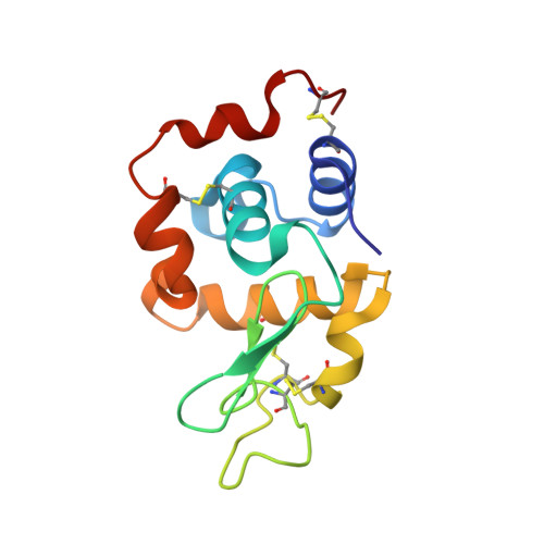
6QXX | pdb_00006qxx

Pink beam serial crystallography: Lysozyme, 5 us exposure, 14793 patterns merged

  • Classification: HYDROLASE
  • Organism(s): Gallus gallus
  • Mutation(s): No 

  • Deposited: 2019-03-08 Released: 2019-10-16 
  • Deposition Author(s): Lieske, J., Tolstikova, A., Meents, A.
  • Funding Organization(s): European Commission, German Federal Ministry for Education and Research, European Union, European Research Council

Experimental Data Snapshot

  • Method: X-RAY DIFFRACTION
  • Resolution: 1.70 Å
  • R-Value Free: 
    0.180 (Depositor), 0.180 (DCC) 
  • R-Value Work: 
    0.151 (Depositor), 0.152 (DCC) 
  • R-Value Observed: 
    0.154 (Depositor) 

Starting Model: experimental
View more details

wwPDB Validation 3D Report Full Report

Validation slider image for 6QXX

This is version 1.2 of the entry. See complete history. 

Literature

1 kHz fixed-target serial crystallography using a multilayer monochromator and an integrating pixel detector.

Tolstikova, A., Levantino, M., Yefanov, O., Hennicke, V., Fischer, P., Meyer, J., Mozzanica, A., Redford, S., Crosas, E., Opara, N.L., Barthelmess, M., Lieske, J., Oberthuer, D., Wator, E., Mohacsi, I., Wulff, M., Schmitt, B., Chapman, H.N., Meents, A.

(2019) IUCrJ 6: 927-937

  • DOI: https://doi.org/10.1107/S205225251900914X
  • Primary Citation Related Structures: 
    6QXV, 6QXW, 6QXX, 6QXY, 6QY0, 6QY1, 6QY2, 6QY4, 6QY5

  • PubMed Abstract: 

    Reliable sample delivery and efficient use of limited beam time have remained bottlenecks for serial crystallography (SX). Using a high-intensity polychromatic X-ray beam in combination with a newly developed charge-integrating JUNGFRAU detector, we have applied the method of fixed-target SX to collect data at a rate of 1 kHz at a synchrotron-radiation facility. According to our data analysis for the given experimental conditions, only about 3 000 diffraction patterns are required for a high-quality diffraction dataset. With indexing rates of up to 25%, recording of such a dataset takes less than 30 s.


  • Organizational Affiliation: 
    • Center for Free Electron Laser Science, DESY, Notkestrasse 85, 22607 Hamburg, Germany.

Macromolecule Content 

  • Total Structure Weight: 14.51 kDa 
  • Atom Count: 1,166 
  • Modeled Residue Count: 129 
  • Deposited Residue Count: 129 
  • Unique protein chains: 1

Macromolecules

Find similar proteins by:|  3D Structure
Entity ID: 1
MoleculeChains  Sequence LengthOrganismDetailsImage
Lysozyme C129Gallus gallusMutation(s): 0 
EC: 3.2.1.17
UniProt
Find proteins for P00698 (Gallus gallus)
Explore P00698 
Go to UniProtKB:  P00698
Entity Groups
Sequence Clusters30% Identity50% Identity70% Identity90% Identity95% Identity100% Identity
UniProt GroupP00698
Sequence Annotations
Expand
Reference Sequence

Experimental Data & Validation

Experimental Data

  • Method: X-RAY DIFFRACTION
  • Resolution: 1.70 Å
  • R-Value Free:  0.180 (Depositor), 0.180 (DCC) 
  • R-Value Work:  0.151 (Depositor), 0.152 (DCC) 
  • R-Value Observed: 0.154 (Depositor) 
Space Group: P 43 21 2
Unit Cell:
Length ( Å )Angle ( ˚ )
a = 79.86α = 90
b = 79.86β = 90
c = 38.02γ = 90
Software Package:
Software NamePurpose
PHENIXrefinement
PDB_EXTRACTdata extraction
CrystFELdata reduction
CrystFELdata scaling
PHASERphasing
Cootmodel building

Structure Validation

View Full Validation Report


Entry History 

& Funding Information

Deposition Data


Funding OrganizationLocationGrant Number
European Commission637295
German Federal Ministry for Education and ResearchGermany031B0405D
European Union654220
European Research Council609920
German Federal Ministry for Education and ResearchGermany05K2018-201-06727 MXD

Revision History  (Full details and data files)

  • Version 1.0: 2019-10-16
    Type: Initial release
  • Version 1.1: 2024-01-24
    Changes: Data collection, Database references, Refinement description
  • Version 1.2: 2024-10-23
    Changes: Structure summary