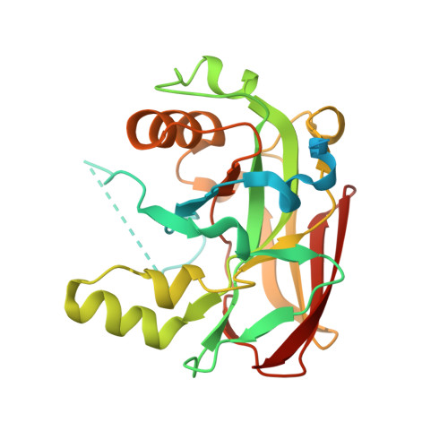
6SBJ

X-ray structure of mus musculus Fumarylacetoacetate hydrolase domain containing protein 1 (FAHD1) apo-form uuncomplexed


Experimental Data Snapshot

  • Method: X-RAY DIFFRACTION
  • Resolution: 2.22 Å
  • R-Value Free: 0.257 
  • R-Value Work: 0.193 
  • R-Value Observed: 0.196 

Starting Model: experimental
View more details

wwPDB Validation   3D Report Full Report


This is version 1.2 of the entry. See complete history


Literature

Structural and functional comparison of fumarylacetoacetate domain containing protein 1 in human and mouse.

Weiss, A.K.H.Naschberger, A.Cappuccio, E.Metzger, C.Mottes, L.Holzknecht, M.von Velsen, J.Bowler, M.W.Rupp, B.Jansen-Durr, P.

(2020) Biosci Rep 40

  • DOI: https://doi.org/10.1042/BSR20194431
  • Primary Citation of Related Structures:  
    6SBI, 6SBJ

  • PubMed Abstract: 

    FAH domain containing protein 1 (FAHD1) is a mammalian mitochondrial protein, displaying bifunctionality as acylpyruvate hydrolase (ApH) and oxaloacetate decarboxylase (ODx) activity. We report the crystal structure of mouse FAHD1 and structural mapping of the active site of mouse FAHD1. Despite high structural similarity with human FAHD1, a rabbit monoclonal antibody (RabMab) could be produced that is able to recognize mouse FAHD1, but not the human form, whereas a polyclonal antibody recognized both proteins. Epitope mapping in combination with our deposited crystal structures revealed that the epitope overlaps with a reported SIRT3 deacetylation site in mouse FAHD1.


  • Organizational Affiliation

    University of Innsbruck, Research Institute for Biomedical Aging Research, Rennweg 10, Innsbruck A-6020, Austria.


Macromolecules
Find similar proteins by:  (by identity cutoff)  |  3D Structure
Entity ID: 1
MoleculeChains Sequence LengthOrganismDetailsImage
Acylpyruvase FAHD1, mitochondrial
A, B, C, D
265Mus musculusMutation(s): 0 
Gene Names: Fahd1MNCb-4134
EC: 3.7.1.5 (PDB Primary Data), 4.1.1.112 (PDB Primary Data), 5.3.2.2 (UniProt)
UniProt
Find proteins for Q8R0F8 (Mus musculus)
Explore Q8R0F8 
Go to UniProtKB:  Q8R0F8
Entity Groups  
Sequence Clusters30% Identity50% Identity70% Identity90% Identity95% Identity100% Identity
UniProt GroupQ8R0F8
Sequence Annotations
Expand
  • Reference Sequence
Small Molecules
Ligands 2 Unique
IDChains Name / Formula / InChI Key2D Diagram3D Interactions
CL
Query on CL

Download Ideal Coordinates CCD File 
G [auth A]
H [auth A]
J [auth B]
L [auth C]
M [auth C]
G [auth A],
H [auth A],
J [auth B],
L [auth C],
M [auth C],
P [auth D]
CHLORIDE ION
Cl
VEXZGXHMUGYJMC-UHFFFAOYSA-M
MG
Query on MG

Download Ideal Coordinates CCD File 
E [auth A]
F [auth A]
I [auth B]
K [auth C]
N [auth D]
E [auth A],
F [auth A],
I [auth B],
K [auth C],
N [auth D],
O [auth D]
MAGNESIUM ION
Mg
JLVVSXFLKOJNIY-UHFFFAOYSA-N
Experimental Data & Validation

Experimental Data

Unit Cell:
Length ( Å )Angle ( ˚ )
a = 52.401α = 90
b = 69.959β = 92.25
c = 107.557γ = 90
Software Package:
Software NamePurpose
REFMACrefinement
PDB_EXTRACTdata extraction
XDSdata reduction
STARANISOdata scaling
MOLREPphasing

Structure Validation

View Full Validation Report


Entry History & Funding Information

Deposition Data


Funding OrganizationLocationGrant Number
European CommissionAustria512020
Austrian Science FundAustriaP28395-B26

Revision History  (Full details and data files)

  • Version 1.0: 2020-02-26
    Type: Initial release
  • Version 1.1: 2020-03-11
    Changes: Database references
  • Version 1.2: 2024-01-24
    Changes: Data collection, Database references, Derived calculations, Refinement description