6T79

Structure of a human nucleosome at 3.2 A resolution


Experimental Data Snapshot

  • Method: ELECTRON MICROSCOPY
  • Resolution: 3.20 Å
  • Aggregation State: PARTICLE 
  • Reconstruction Method: SINGLE PARTICLE 

Starting Model: experimental
View more details

wwPDB Validation   3D Report Full Report

Currently 6T79 does not have a validation slider image.


This is version 1.2 of the entry. See complete history


Literature

Nucleosome-bound SOX2 and SOX11 structures elucidate pioneer factor function.

Dodonova, S.O.Zhu, F.Dienemann, C.Taipale, J.Cramer, P.

(2020) Nature 580: 669-672

  • DOI: https://doi.org/10.1038/s41586-020-2195-y
  • Primary Citation of Related Structures:  
    6T78, 6T79, 6T7A, 6T7B, 6T7C, 6T7D

  • PubMed Abstract: 

    'Pioneer' transcription factors are required for stem-cell pluripotency, cell differentiation and cell reprogramming 1,2 . Pioneer factors can bind nucleosomal DNA to enable gene expression from regions of the genome with closed chromatin. SOX2 is a prominent pioneer factor that is essential for pluripotency and self-renewal of embryonic stem cells 3 . Here we report cryo-electron microscopy structures of the DNA-binding domains of SOX2 and its close homologue SOX11 bound to nucleosomes. The structures show that SOX factors can bind and locally distort DNA at superhelical location 2. The factors also facilitate detachment of terminal nucleosomal DNA from the histone octamer, which increases DNA accessibility. SOX-factor binding to the nucleosome can also lead to a repositioning of the N-terminal tail of histone H4 that includes residue lysine 16. We speculate that this repositioning is incompatible with higher-order nucleosome stacking, which involves contacts of the H4 tail with a neighbouring nucleosome. Our results indicate that pioneer transcription factors can use binding energy to initiate chromatin opening, and thereby facilitate nucleosome remodelling and subsequent transcription.


  • Organizational Affiliation

    Department of Molecular Biology, Max Planck Institute for Biophysical Chemistry, Göttingen, Germany.


Macromolecules

Find similar proteins by:  (by identity cutoff)  |  3D Structure
Entity ID: 1
MoleculeChains Sequence LengthOrganismDetailsImage
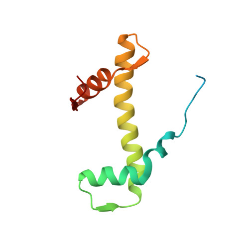
Histone H3.2
A, E
136Homo sapiensMutation(s): 1 
Gene Names: HIST2H3AHIST2H3CH3F2H3FMHIST2H3D
UniProt & NIH Common Fund Data Resources
Find proteins for Q71DI3 (Homo sapiens)
Go to UniProtKB:  Q71DI3
PHAROS:  Q71DI3
Sequence Annotations
Expand
  • Reference Sequence
Find similar proteins by:  (by identity cutoff)  |  3D Structure
Entity ID: 2
MoleculeChains Sequence LengthOrganismDetailsImage
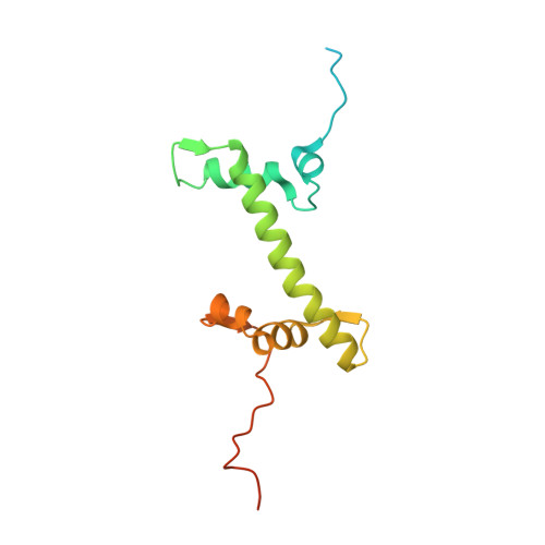
Histone H4
B, F
103Homo sapiensMutation(s): 0 
Gene Names: 
UniProt & NIH Common Fund Data Resources
Find proteins for P62805 (Homo sapiens)
Go to UniProtKB:  P62805
PHAROS:  P62805
Sequence Annotations
Expand
  • Reference Sequence
Find similar proteins by:  (by identity cutoff)  |  3D Structure
Entity ID: 3
MoleculeChains Sequence LengthOrganismDetailsImage
Histone H2A type 1-B/E
C, G
151Homo sapiensMutation(s): 0 
Gene Names: HIST1H2ABH2AFMHIST1H2AEH2AFA
UniProt & NIH Common Fund Data Resources
Find proteins for P04908 (Homo sapiens)
Go to UniProtKB:  P04908
PHAROS:  P04908
Sequence Annotations
Expand
  • Reference Sequence
Find similar proteins by:  (by identity cutoff)  |  3D Structure
Entity ID: 4
MoleculeChains Sequence LengthOrganismDetailsImage
Histone H2B type 1-K
D, H
126Homo sapiensMutation(s): 0 
Gene Names: HIST1H2BKH2BFTHIRIP1
UniProt & NIH Common Fund Data Resources
Find proteins for O60814 (Homo sapiens)
Go to UniProtKB:  O60814
PHAROS:  O60814
GTEx:  ENSG00000197903 
Sequence Annotations
Expand
  • Reference Sequence
Find similar nucleic acids by:  (by identity cutoff)  |  3D Structure
Entity ID: 5
MoleculeChains LengthOrganismImage
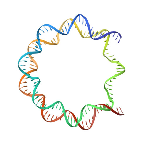
DNA (147-MER)147synthetic construct
Sequence Annotations
Expand
  • Reference Sequence
Find similar nucleic acids by:  (by identity cutoff)  |  3D Structure
Entity ID: 6
MoleculeChains LengthOrganismImage
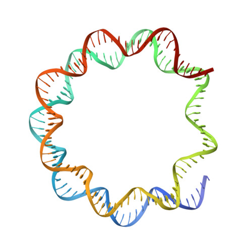
DNA (147-MER)147synthetic construct
Sequence Annotations
Expand
  • Reference Sequence
Experimental Data & Validation

Experimental Data

  • Method: ELECTRON MICROSCOPY
  • Resolution: 3.20 Å
  • Aggregation State: PARTICLE 
  • Reconstruction Method: SINGLE PARTICLE 
EM Software:
TaskSoftware PackageVersion
RECONSTRUCTIONRELION
MODEL REFINEMENTPHENIX

Structure Validation

View Full Validation Report

Currently 6T79 does not have a validation slider image.


Entry History & Funding Information

Deposition Data


Funding OrganizationLocationGrant Number
European Research CouncilGermany693023
European Molecular Biology OrganizationGermanyALTF-949-2016
Volkswagen FoundationGermany--

Revision History  (Full details and data files)

  • Version 1.0: 2020-04-29
    Type: Initial release
  • Version 1.1: 2020-05-13
    Changes: Database references
  • Version 1.2: 2024-05-22
    Changes: Data collection, Database references, Refinement description