6TSA | pdb_00006tsa

Crystal structure of human L ferritin (HuLf) Fe(III)-loaded for 30 minutes


Experimental Data Snapshot

  • Method: X-RAY DIFFRACTION
  • Resolution: 2.18 Å
  • R-Value Free: 
    0.258 (Depositor), 0.258 (DCC) 
  • R-Value Work: 
    0.202 (Depositor), 0.202 (DCC) 

Starting Model: experimental
View more details

wwPDB Validation 3D Report Full Report

Validation slider image for 6TSA

This is version 1.2 of the entry. See complete history

Literature

Iron Biomineral Growth from the Initial Nucleation Seed in L-Ferritin.

Ciambellotti, S.Pozzi, C.Mangani, S.Turano, P.

(2020) Chemistry 26: 5770-5773

  • DOI: https://doi.org/10.1002/chem.202000064
  • Primary Citation Related Structures: 
    6TR9, 6TRZ, 6TS0, 6TS1, 6TSA, 6TSF, 6TSJ, 6TSS, 6TSX

  • PubMed Abstract: 

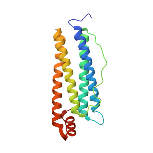
    X-ray structures of homopolymeric human L-ferritin and horse spleen ferritin were solved by freezing protein crystals at different time intervals after exposure to a ferric salt and revealed the growth of an octa-nuclear iron cluster on the inner surface of the protein cage with a key role played by some glutamate residues. An atomic resolution view of how the cluster formation develops starting from a (μ 3 -oxo)tris[(μ 2 -glutamato-κO:κO')](glutamato-κO)(diaquo)triiron(III) seed is provided. The results support the idea that iron biomineralization in ferritin is a process initiating at the level of the protein surface, capable of contributing coordination bonds and electrostatic guidance.


  • Organizational Affiliation
    • Magnetic Resonance Center (CERM), University of Florence, Sesto Fiorentino, 50019, Italy.

Macromolecule Content 

  • Total Structure Weight: 21.06 kDa 
  • Atom Count: 1,566 
  • Modeled Residue Count: 172 
  • Deposited Residue Count: 175 
  • Unique protein chains: 1

Macromolecules

Find similar proteins by:|  3D Structure
Entity ID: 1
MoleculeChains  Sequence LengthOrganismDetailsImage
Ferritin light chainA [auth AAA]175Homo sapiensMutation(s): 0 
Gene Names: FTL
UniProt & NIH Common Fund Data Resources
Find proteins for P02792 (Homo sapiens)
Explore P02792 
Go to UniProtKB:  P02792
PHAROS:  P02792
GTEx:  ENSG00000087086 
Entity Groups
Sequence Clusters30% Identity50% Identity70% Identity90% Identity95% Identity100% Identity
UniProt GroupP02792
Sequence Annotations
Expand
Reference Sequence

Small Molecules

Ligands 3 Unique
IDChains Name / Formula / InChI Key2D Diagram3D Interactions
CD

Query on CD


Download:Ideal Coordinates CCD File
Q [auth AAA]CADMIUM ION
Cd
WLZRMCYVCSSEQC-UHFFFAOYSA-N
FE
(Subject of Investigation/LOI)

Query on FE


Download:Ideal Coordinates CCD File
B [auth AAA]
C [auth AAA]
D [auth AAA]
E [auth AAA]
F [auth AAA]
B [auth AAA],
C [auth AAA],
D [auth AAA],
E [auth AAA],
F [auth AAA],
G [auth AAA],
H [auth AAA],
I [auth AAA],
J [auth AAA],
K [auth AAA],
L [auth AAA],
M [auth AAA],
N [auth AAA],
O [auth AAA],
P [auth AAA]
FE (III) ION
Fe
VTLYFUHAOXGGBS-UHFFFAOYSA-N
O

Query on O


Download:Ideal Coordinates CCD File
R [auth AAA],
S [auth AAA],
T [auth AAA],
U [auth AAA]
OXYGEN ATOM
O
XLYOFNOQVPJJNP-UHFFFAOYSA-N

Experimental Data & Validation

Experimental Data

  • Method: X-RAY DIFFRACTION
  • Resolution: 2.18 Å
  • R-Value Free:  0.258 (Depositor), 0.258 (DCC) 
  • R-Value Work:  0.202 (Depositor), 0.202 (DCC) 
Space Group: I 4 3 2
Unit Cell:
Length ( Å )Angle ( ˚ )
a = 152.69α = 90
b = 152.69β = 90
c = 152.69γ = 90
Software Package:
Software NamePurpose
REFMACrefinement
XDSdata reduction
SCALAdata scaling
MOLREPphasing

Structure Validation

View Full Validation Report


Entry History 

Deposition Data

Revision History  (Full details and data files)

  • Version 1.0: 2020-02-19
    Type: Initial release
  • Version 1.1: 2020-05-20
    Changes: Database references
  • Version 1.2: 2024-01-24
    Changes: Data collection, Database references, Derived calculations, Refinement description