6U3E | pdb_00006u3e

Best fitting antiparallel model for Volume 1 of truncated dimeric Cytohesin-3 (Grp1; amino acids 14-399)


Experimental Data Snapshot

  • Method: ELECTRON MICROSCOPY
  • Resolution: 53.0 Å
  • Aggregation State: PARTICLE 
  • Reconstruction Method: SINGLE PARTICLE 

Starting Model: experimental
View more details

wwPDB Validation 3D Report Full Report

Validation slider image for 6U3E

This is version 1.4 of the entry. See complete history

Literature

Structural Organization and Dynamics of Homodimeric Cytohesin Family Arf GTPase Exchange Factors in Solution and on Membranes.

Das, S.Malaby, A.W.Nawrotek, A.Zhang, W.Zeghouf, M.Maslen, S.Skehel, M.Chakravarthy, S.Irving, T.C.Bilsel, O.Cherfils, J.Lambright, D.G.

(2019) Structure 27: 1782

  • DOI: https://doi.org/10.1016/j.str.2019.09.007
  • Primary Citation Related Structures: 
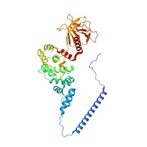
    6U3E, 6U3G

  • PubMed Abstract: 

    Membrane dynamic processes require Arf GTPase activation by guanine nucleotide exchange factors (GEFs) with a Sec7 domain. Cytohesin family Arf GEFs function in signaling and cell migration through Arf GTPase activation on the plasma membrane and endosomes. In this study, the structural organization of two cytohesins (Grp1 and ARNO) was investigated in solution by size exclusion-small angle X-ray scattering and negative stain-electron microscopy and on membranes by dynamic light scattering, hydrogen-deuterium exchange-mass spectrometry and guanosine diphosphate (GDP)/guanosine triphosphate (GTP) exchange assays. The results suggest that cytohesins form elongated dimers with a central coiled coil and membrane-binding pleckstrin-homology (PH) domains at opposite ends. The dimers display significant conformational heterogeneity, with a preference for compact to intermediate conformations. Phosphoinositide-dependent membrane recruitment is mediated by one PH domain at a time and alters the conformational dynamics to prime allosteric activation by Arf-GTP. A structural model for membrane targeting and allosteric activation of full-length cytohesin dimers is discussed.


  • Organizational Affiliation
    • Program in Molecular Medicine, University of Massachusetts Medical School, Worcester, MA 01605, USA.

Macromolecule Content 

  • Total Structure Weight: 94 kDa 
  • Atom Count: 6,594 
  • Modeled Residue Count: 794 
  • Deposited Residue Count: 794 
  • Unique protein chains: 1

Macromolecules

Find similar proteins by:|  3D Structure
Entity ID: 1
MoleculeChains  Sequence LengthOrganismDetailsImage
Cytohesin-3
A, B
397Homo sapiensMutation(s): 0 
Gene Names: CYTH3ARNO3GRP1PSCD3
UniProt & NIH Common Fund Data Resources
Find proteins for O43739 (Homo sapiens)
Explore O43739 
Go to UniProtKB:  O43739
PHAROS:  O43739
GTEx:  ENSG00000008256 
Entity Groups
Sequence Clusters30% Identity50% Identity70% Identity90% Identity95% Identity100% Identity
UniProt GroupO43739
Sequence Annotations
Expand
Reference Sequence

Small Molecules

Binding Affinity Annotations 
IDSourceBinding Affinity
4IP BindingDB:  6U3E Kd: 100 (nM) from 1 assay(s)

Experimental Data & Validation

Experimental Data

  • Method: ELECTRON MICROSCOPY
  • Resolution: 53.0 Å
  • Aggregation State: PARTICLE 
  • Reconstruction Method: SINGLE PARTICLE 
EM Software:
TaskSoftware PackageVersion
RECONSTRUCTIONEMAN22.12

Structure Validation

View Full Validation Report


Entry History 

& Funding Information

Deposition Data


Funding OrganizationLocationGrant Number
National Institutes of Health/National Institute of General Medical Sciences (NIH/NIGMS)United StatesGM056324

Revision History  (Full details and data files)

  • Version 1.0: 2019-09-25
    Type: Initial release
  • Version 1.1: 2019-10-23
    Changes: Data collection, Database references
  • Version 1.2: 2019-12-18
    Changes: Database references
  • Version 1.3: 2020-01-08
    Changes: Author supporting evidence
  • Version 1.4: 2023-11-29
    Changes: Data collection, Database references, Refinement description