6UIL

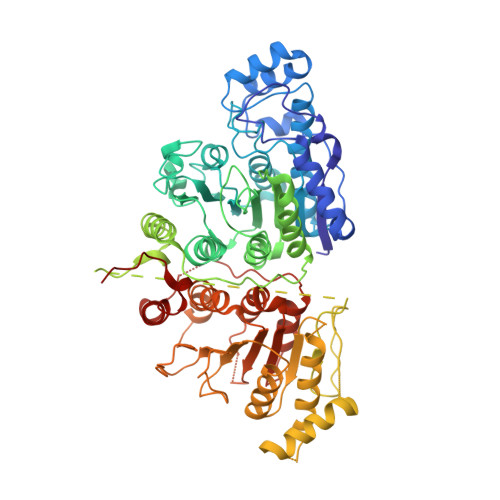
Crystal Structure of Danio rerio Histone Deacetylase 10 in Complex with 7-[(3-aminopropyl)amino]-1,1,1-trifluoroheptan-2-one


Experimental Data Snapshot

  • Method: X-RAY DIFFRACTION
  • Resolution: 2.85 Å
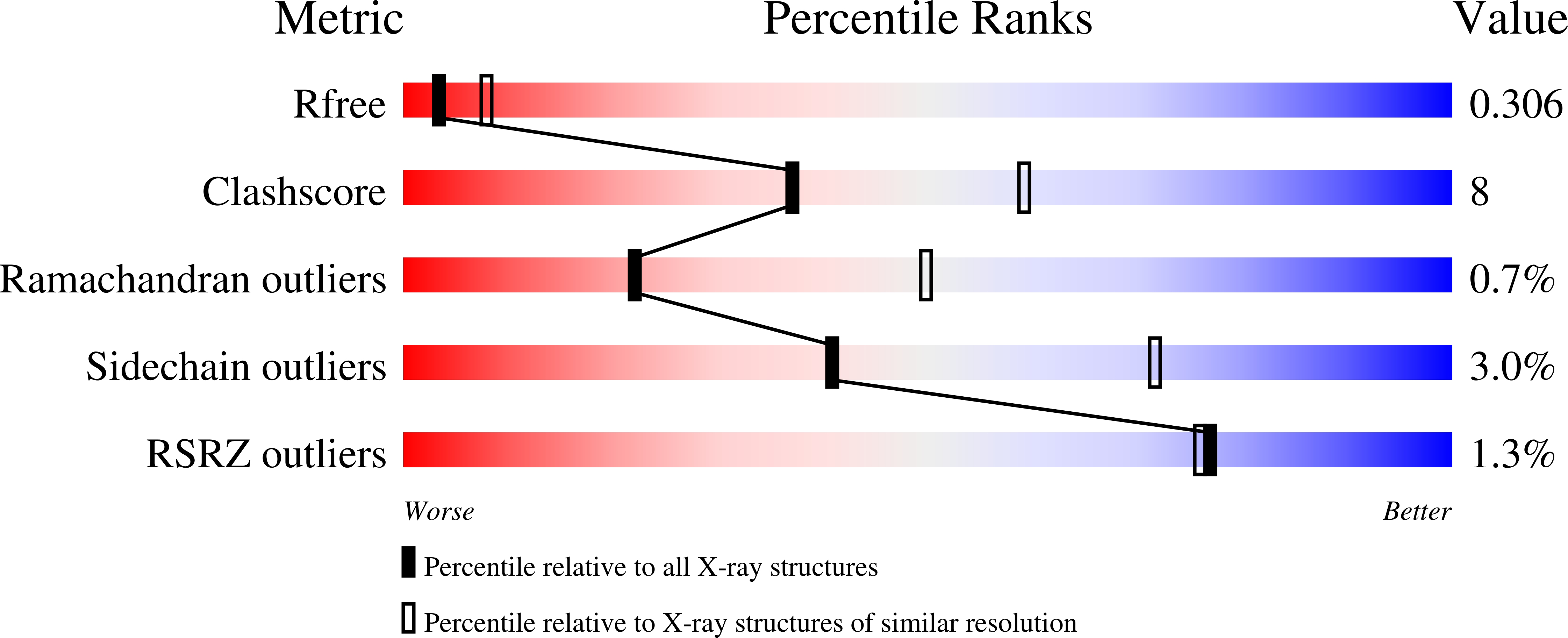
  • R-Value Free: 0.306 
  • R-Value Work: 0.244 
  • R-Value Observed: 0.247 

Starting Model: experimental
View more details

wwPDB Validation   3D Report Full Report


Ligand Structure Quality Assessment 


This is version 1.2 of the entry. See complete history


Literature

Binding ofN8-Acetylspermidine Analogues to Histone Deacetylase 10 Reveals Molecular Strategies for Blocking Polyamine Deacetylation.

Herbst-Gervasoni, C.J.Christianson, D.W.

(2019) Biochemistry 58: 4957-4969

  • DOI: https://doi.org/10.1021/acs.biochem.9b00906
  • Primary Citation of Related Structures:  
    6UFN, 6UFO, 6UHU, 6UHV, 6UII, 6UIJ, 6UIL, 6UIM

  • PubMed Abstract: 

    Eukaryotic histone deacetylase 10 (HDAC10) is a Zn 2+ -dependent hydrolase that exhibits catalytic specificity for the hydrolysis of the polyamine N 8 -acetylspermidine. The recently determined crystal structure of HDAC10 from Danio rerio (zebrafish) reveals a narrow active site cleft and a negatively charged "gatekeeper" (E274) that favors the binding of the slender cationic substrate. Because HDAC10 expression is upregulated in advanced-stage neuroblastoma and induces autophagy, the selective inhibition of HDAC10 suppresses the autophagic response and renders cancer cells more susceptible to cytotoxic chemotherapeutic drugs. Here, we describe X-ray crystal structures of zebrafish HDAC10 complexed with eight different analogues of N 8 -acetylspermidine. These analogues contain different Zn 2+ -binding groups, such as hydroxamate, thiolate, and the tetrahedral gem-diolate resulting from the addition of a Zn 2+ -bound water molecule to a ketone carbonyl group. Notably, the chemistry that accompanies the binding of ketonic substrate analogues is identical to the chemistry involved in the first step of catalysis, i.e., nucleophilic attack of a Zn 2+ -bound water molecule at the scissile carbonyl group of N 8 -acetylspermidine. The most potent inhibitor studied contains a thiolate Zn 2+ -binding group. These structures reveal interesting geometric changes in the metal coordination polyhedron that accommodate inhibitor binding. Additional interactions in the active site highlight features contributing to substrate specificity. These interactions are likely to contribute to inhibitor binding selectivity and will inform the future design of compounds selective for HDAC10 inhibition.


  • Organizational Affiliation

    Roy and Diana Vagelos Laboratories, Department of Chemistry , University of Pennsylvania , Philadelphia , Pennsylvania 19104-6323 , United States.


Macromolecules
Find similar proteins by:  (by identity cutoff)  |  3D Structure
Entity ID: 1
MoleculeChains Sequence LengthOrganismDetailsImage
Polyamine deacetylase HDAC10676Danio rerioMutation(s): 0 
EC: 3.5.1.48 (PDB Primary Data), 3.5.1.62 (PDB Primary Data)
UniProt
Find proteins for F1QCV2 (Danio rerio)
Explore F1QCV2 
Go to UniProtKB:  F1QCV2
Entity Groups  
Sequence Clusters30% Identity50% Identity70% Identity90% Identity95% Identity100% Identity
UniProt GroupF1QCV2
Sequence Annotations
Expand
  • Reference Sequence
Small Molecules
Ligands 4 Unique
IDChains Name / Formula / InChI Key2D Diagram3D Interactions
FKS (Subject of Investigation/LOI)
Query on FKS

Download Ideal Coordinates CCD File 
B [auth A]7-[(3-aminopropyl)amino]-1,1,1-trifluoroheptane-2,2-diol
C10 H21 F3 N2 O2
IVRAKANOVJJYCX-UHFFFAOYSA-N
PO4
Query on PO4

Download Ideal Coordinates CCD File 
C [auth A],
D [auth A]
PHOSPHATE ION
O4 P
NBIIXXVUZAFLBC-UHFFFAOYSA-K
ZN
Query on ZN

Download Ideal Coordinates CCD File 
E [auth A]ZINC ION
Zn
PTFCDOFLOPIGGS-UHFFFAOYSA-N
K
Query on K

Download Ideal Coordinates CCD File 
F [auth A],
G [auth A]
POTASSIUM ION
K
NPYPAHLBTDXSSS-UHFFFAOYSA-N
Experimental Data & Validation

Experimental Data

  • Method: X-RAY DIFFRACTION
  • Resolution: 2.85 Å
  • R-Value Free: 0.306 
  • R-Value Work: 0.244 
  • R-Value Observed: 0.247 
  • Space Group: P 31 2 1
Unit Cell:
Length ( Å )Angle ( ˚ )
a = 80.98α = 90
b = 80.98β = 90
c = 242.822γ = 120
Software Package:
Software NamePurpose
PHENIXrefinement
iMOSFLMdata reduction
Aimlessdata scaling
PHASERphasing

Structure Validation

View Full Validation Report


Ligand Structure Quality Assessment 


Entry History & Funding Information

Deposition Data


Funding OrganizationLocationGrant Number
National Institutes of Health/National Human Genome Research Institute (NIH/NHGRI)United StatesGM49758

Revision History  (Full details and data files)

  • Version 1.0: 2019-12-04
    Type: Initial release
  • Version 1.1: 2019-12-18
    Changes: Database references
  • Version 1.2: 2023-10-11
    Changes: Data collection, Database references, Derived calculations, Refinement description