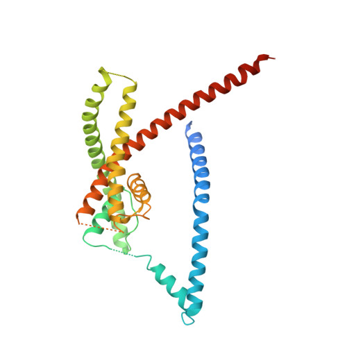
6V36

K2P2.1(TREK-1)I110D apo channel structure


Experimental Data Snapshot

  • Method: X-RAY DIFFRACTION
  • Resolution: 3.40 Å
  • R-Value Free: 0.318 
  • R-Value Work: 0.286 
  • R-Value Observed: 0.288 

Starting Model: experimental
View more details

wwPDB Validation   3D Report Full Report

Currently 6V36 does not have a validation slider image.


This is version 1.2 of the entry. See complete history


Literature

Polynuclear Ruthenium Amines Inhibit K2PChannels via a "Finger in the Dam" Mechanism.

Pope, L.Lolicato, M.Minor Jr., D.L.

(2020) Cell Chem Biol 

  • DOI: https://doi.org/10.1016/j.chembiol.2020.01.011
  • Primary Citation of Related Structures:  
    6V36, 6V37, 6V3C, 6V3I

  • PubMed Abstract: 

    The trinuclear ruthenium amine ruthenium red (RuR) inhibits diverse ion channels, including K 2P potassium channels, TRPs, the calcium uniporter, CALHMs, ryanodine receptors, and Piezos. Despite this extraordinary array, there is limited information for how RuR engages targets. Here, using X-ray crystallographic and electrophysiological studies of an RuR-sensitive K 2P , K 2P 2.1 (TREK-1) I110D, we show that RuR acts by binding an acidic residue pair comprising the "Keystone inhibitor site" under the K 2P CAP domain archway above the channel pore. We further establish that Ru360, a dinuclear ruthenium amine not known to affect K 2P s, inhibits RuR-sensitive K 2P s using the same mechanism. Structural knowledge enabled a generalizable design strategy for creating K 2P RuR "super-responders" having nanomolar sensitivity. Together, the data define a "finger in the dam" inhibition mechanism acting at a novel K 2P inhibitor binding site. These findings highlight the polysite nature of K 2P pharmacology and provide a new framework for K 2P inhibitor development.


  • Organizational Affiliation

    Cardiovascular Research Institute, University of California, San Francisco, CA 93858-2330, USA.


Macromolecules
Find similar proteins by:  (by identity cutoff)  |  3D Structure
Entity ID: 1
MoleculeChains Sequence LengthOrganismDetailsImage
Potassium channel subfamily K member 2
A, B
312Mus musculusMutation(s): 0 
Membrane Entity: Yes 
UniProt & NIH Common Fund Data Resources
Find proteins for P97438 (Mus musculus)
Explore P97438 
Go to UniProtKB:  P97438
IMPC:  MGI:109366
Entity Groups  
Sequence Clusters30% Identity50% Identity70% Identity90% Identity95% Identity100% Identity
UniProt GroupP97438
Sequence Annotations
Expand
  • Reference Sequence
Small Molecules
Ligands 4 Unique
IDChains Name / Formula / InChI Key2D Diagram3D Interactions
UND
Query on UND

Download Ideal Coordinates CCD File 
F [auth A],
O [auth B]
UNDECANE
C11 H24
RSJKGSCJYJTIGS-UHFFFAOYSA-N
OCT
Query on OCT

Download Ideal Coordinates CCD File 
D [auth A],
E [auth A],
N [auth B]
N-OCTANE
C8 H18
TVMXDCGIABBOFY-UHFFFAOYSA-N
CD
Query on CD

Download Ideal Coordinates CCD File 
C [auth A],
L [auth B],
M [auth B]
CADMIUM ION
Cd
WLZRMCYVCSSEQC-UHFFFAOYSA-N
K
Query on K

Download Ideal Coordinates CCD File 
G [auth A]
H [auth A]
I [auth A]
J [auth A]
K [auth A]
G [auth A],
H [auth A],
I [auth A],
J [auth A],
K [auth A],
P [auth B]
POTASSIUM ION
K
NPYPAHLBTDXSSS-UHFFFAOYSA-N
Experimental Data & Validation

Experimental Data

  • Method: X-RAY DIFFRACTION
  • Resolution: 3.40 Å
  • R-Value Free: 0.318 
  • R-Value Work: 0.286 
  • R-Value Observed: 0.288 
  • Space Group: P 21 21 21
Unit Cell:
Length ( Å )Angle ( ˚ )
a = 68.192α = 90
b = 120.4β = 90
c = 128.355γ = 90
Software Package:
Software NamePurpose
PHENIXrefinement
XDSdata reduction
Aimlessdata scaling
PHASERphasing

Structure Validation

View Full Validation Report

Currently 6V36 does not have a validation slider image.


Entry History & Funding Information

Deposition Data


Funding OrganizationLocationGrant Number
National Institutes of Health/National Library of Medicine (NIH/NLM)United StatesNIH-R01-MH093603

Revision History  (Full details and data files)

  • Version 1.0: 2020-02-26
    Type: Initial release
  • Version 1.1: 2023-10-11
    Changes: Data collection, Database references, Derived calculations, Refinement description
  • Version 1.2: 2024-11-20
    Changes: Structure summary