6XF7 | pdb_00006xf7

SLP


Experimental Data Snapshot

  • Method: ELECTRON MICROSCOPY
  • Resolution: 6.60 Å
  • Aggregation State: CELL 
  • Reconstruction Method: SUBTOMOGRAM AVERAGING 

wwPDB Validation 3D Report Full Report

Validation slider image for 6XF7

This is version 1.1 of the entry. See complete history

Literature

Assembly intermediates of orthoreovirus captured in the cell.

Sutton, G.Sun, D.Fu, X.Kotecha, A.Hecksel, C.W.Clare, D.K.Zhang, P.Stuart, D.I.Boyce, M.

(2020) Nat Commun 11: 4445-4445

  • DOI: https://doi.org/10.1038/s41467-020-18243-9
  • Primary Citation Related Structures: 
    6XF7, 6XF8, 6ZTS, 6ZTY, 6ZTZ

  • PubMed Abstract: 

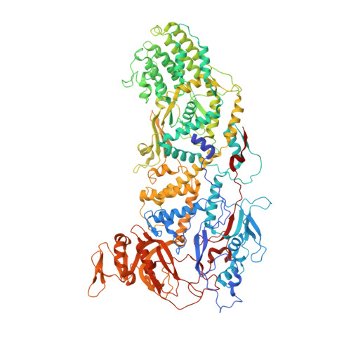
    Traditionally, molecular assembly pathways for viruses are inferred from high resolution structures of purified stable intermediates, low resolution images of cell sections and genetic approaches. Here, we directly visualise an unsuspected 'single shelled' intermediate for a mammalian orthoreovirus in cryo-preserved infected cells, by cryo-electron tomography of cellular lamellae. Particle classification and averaging yields structures to 5.6 Å resolution, sufficient to identify secondary structural elements and produce an atomic model of the intermediate, comprising 120 copies each of protein λ1 and σ2. This λ1 shell is 'collapsed' compared to the mature virions, with molecules pushed inwards at the icosahedral fivefolds by ~100 Å, reminiscent of the first assembly intermediate of certain prokaryotic dsRNA viruses. This supports the supposition that these viruses share a common ancestor, and suggests mechanisms for the assembly of viruses of the Reoviridae. Such methodology holds promise for dissecting the replication cycle of many viruses.


  • Organizational Affiliation
    • Division of Structural Biology, Wellcome Centre for Human Genetics, University of Oxford, Oxford, OX3 7BN, UK.

Macromolecule Content 

  • Total Structure Weight: 238.04 kDa 
  • Atom Count: 16,454 
  • Modeled Residue Count: 2,082 
  • Deposited Residue Count: 2,118 
  • Unique protein chains: 1

Macromolecules

Find similar proteins by:|  3D Structure
Entity ID: 1
MoleculeChains  Sequence LengthOrganismDetailsImage
Lambda 1 proteinA [auth B],
B [auth C]
1,059Mammalian orthoreovirusMutation(s): 0 
EC: 3.6.4.13
UniProt
Find proteins for Q9WAB2 (Reovirus type 1 (strain Lang))
Explore Q9WAB2 
Go to UniProtKB:  Q9WAB2
Entity Groups
Sequence Clusters30% Identity50% Identity70% Identity90% Identity95% Identity100% Identity
UniProt GroupQ9WAB2
Sequence Annotations
Expand
Reference Sequence

Experimental Data & Validation

Experimental Data

  • Method: ELECTRON MICROSCOPY
  • Resolution: 6.60 Å
  • Aggregation State: CELL 
  • Reconstruction Method: SUBTOMOGRAM AVERAGING 
EM Software:
TaskSoftware PackageVersion
RECONSTRUCTIONemClarity

Structure Validation

View Full Validation Report


Entry History 

& Funding Information

Deposition Data


Funding OrganizationLocationGrant Number
Medical Research Council (MRC, United Kingdom)United KingdomMR/N00065X/1
Wellcome TrustUnited Kingdom206422/Z/17/Z
National Institutes of Health/National Institute of Biomedical Imaging and Bioengineering (NIH/NIBIB)United StatesGM082251

Revision History  (Full details and data files)

  • Version 1.0: 2020-09-23
    Type: Initial release
  • Version 1.1: 2024-03-06
    Changes: Data collection, Database references