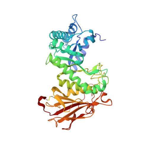
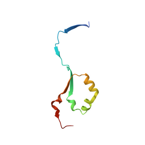
6ZDM

Crystal structure of human heparanase in complex with a N',6O'-bis-sulfated 4-methylumbelliferyl heparan sulfate disaccharide

  • Classification: HYDROLASE
  • Organism(s): Homo sapiens
  • Expression System: Trichoplusia ni
  • Mutation(s): No 

  • Deposited: 2020-06-14 Released: 2020-10-21 
  • Deposition Author(s): Wu, L., Davies, G.J.
  • Funding Organization(s): European Research Council (ERC), Australian Research Council (ARC)

Experimental Data Snapshot

  • Method: X-RAY DIFFRACTION
  • Resolution: 1.71 Å
  • R-Value Free: 0.241 
  • R-Value Work: 0.179 

Starting Model: experimental
View more details

wwPDB Validation   3D Report Full Report


Ligand Structure Quality Assessment 


This is version 1.3 of the entry. See complete history


Literature

Structural insights into heparanase activity using a fluorogenic heparan sulfate disaccharide.

Wu, L.Wimmer, N.Davies, G.J.Ferro, V.

(2020) Chem Commun (Camb) 56: 13780-13783

  • DOI: https://doi.org/10.1039/d0cc05932c
  • Primary Citation of Related Structures:  
    6ZDM

  • PubMed Abstract: 

    A synthetic heparan sulfate disaccharide has been assessed as a fluorogenic heparanase substrate, enabling enzyme turnover and inhibition kinetics measurements despite slow turnover. Crystal structures with human heparanase also provide the first ever observation of a substrate in an activated 1S3 conformation, highlighting previously unknown interactions involved in enzymatic processing. Our data provide insights into the heparanase catalytic mechanism, and will inform the design of improved heparanase substrates and inhibitors.


  • Organizational Affiliation

    Department of Chemistry, University of York, Heslington, York, YO10 5DD, UK. gideon.davies@york.ac.uk.


Macromolecules
Find similar proteins by:  (by identity cutoff)  |  3D Structure
Entity ID: 1
MoleculeChains Sequence LengthOrganismDetailsImage
HeparanaseA [auth AAA]389Homo sapiensMutation(s): 0 
Gene Names: HPSEHEPHPAHPA1HPR1HPSE1HSE1
EC: 3.2.1.166
UniProt & NIH Common Fund Data Resources
Find proteins for Q9Y251 (Homo sapiens)
Explore Q9Y251 
Go to UniProtKB:  Q9Y251
PHAROS:  Q9Y251
GTEx:  ENSG00000173083 
Entity Groups  
Sequence Clusters30% Identity50% Identity70% Identity90% Identity95% Identity100% Identity
UniProt GroupQ9Y251
Glycosylation
Glycosylation Sites: 1Go to GlyGen: Q9Y251-1
Sequence Annotations
Expand
  • Reference Sequence
Find similar proteins by:  (by identity cutoff)  |  3D Structure
Entity ID: 2
MoleculeChains Sequence LengthOrganismDetailsImage
HeparanaseB [auth BBB]77Homo sapiensMutation(s): 0 
Gene Names: HPSEHEPHPAHPA1HPR1HPSE1HSE1
EC: 3.2.1.166
UniProt & NIH Common Fund Data Resources
Find proteins for Q9Y251 (Homo sapiens)
Explore Q9Y251 
Go to UniProtKB:  Q9Y251
PHAROS:  Q9Y251
GTEx:  ENSG00000173083 
Entity Groups  
Sequence Clusters30% Identity50% Identity70% Identity90% Identity95% Identity100% Identity
UniProt GroupQ9Y251
Sequence Annotations
Expand
  • Reference Sequence
Small Molecules
Ligands 4 Unique
IDChains Name / Formula / InChI Key2D Diagram3D Interactions
QG2 (Subject of Investigation/LOI)
Query on QG2

Download Ideal Coordinates CCD File 
E [auth AAA](2~{S},3~{S},4~{R},5~{R},6~{S})-3-[(2~{R},3~{R},4~{R},5~{S},6~{R})-4,5-bis(oxidanyl)-3-(sulfoamino)-6-(sulfooxymethyl)oxan-2-yl]oxy-6-(4-methyl-2-oxidanylidene-chromen-7-yl)oxy-4,5-bis(oxidanyl)oxane-2-carboxylic acid
C22 H27 N O19 S2
VWVHHIREMFENNG-XOAMEYKJSA-N
NAG
Query on NAG

Download Ideal Coordinates CCD File 
C [auth AAA]2-acetamido-2-deoxy-beta-D-glucopyranose
C8 H15 N O6
OVRNDRQMDRJTHS-FMDGEEDCSA-N
SO4
Query on SO4

Download Ideal Coordinates CCD File 
F [auth AAA],
G [auth AAA],
H [auth AAA],
I [auth AAA],
J [auth AAA]
SULFATE ION
O4 S
QAOWNCQODCNURD-UHFFFAOYSA-L
EDO
Query on EDO

Download Ideal Coordinates CCD File 
D [auth AAA]1,2-ETHANEDIOL
C2 H6 O2
LYCAIKOWRPUZTN-UHFFFAOYSA-N
Experimental Data & Validation

Experimental Data

  • Method: X-RAY DIFFRACTION
  • Resolution: 1.71 Å
  • R-Value Free: 0.241 
  • R-Value Work: 0.179 
  • Space Group: P 1 21 1
Unit Cell:
Length ( Å )Angle ( ˚ )
a = 44.974α = 90
b = 70.856β = 97.128
c = 78.455γ = 90
Software Package:
Software NamePurpose
REFMACrefinement
xia2data reduction
STARANISOdata scaling
REFMACphasing

Structure Validation

View Full Validation Report


Ligand Structure Quality Assessment 


Entry History & Funding Information

Deposition Data


Funding OrganizationLocationGrant Number
European Research Council (ERC)United KingdomERC-2012-AdG-322942
Australian Research Council (ARC)AustraliaDP170104431

Revision History  (Full details and data files)

  • Version 1.0: 2020-10-21
    Type: Initial release
  • Version 1.1: 2020-11-04
    Changes: Database references
  • Version 1.2: 2020-11-18
    Changes: Database references
  • Version 1.3: 2024-01-24
    Changes: Data collection, Database references, Derived calculations, Refinement description