7BGF | pdb_00007bgf

CRYSTAL STRUCTURE OF THE N-TERMINAL DIMERIC COILED COIL OF THE HUMAN CTIP PROTEIN


Experimental Data Snapshot

  • Method: X-RAY DIFFRACTION
  • Resolution: 2.80 Å
  • R-Value Free: 
    0.253 (Depositor), 0.256 (DCC) 
  • R-Value Work: 
    0.224 (Depositor), 0.228 (DCC) 

Starting Model: in silico
View more details

wwPDB Validation 3D Report Full Report

Validation slider image for 7BGF

This is version 1.1 of the entry. See complete history

Literature

Structural basis for the coiled-coil architecture of human CtIP.

Morton, C.R.Rzechorzek, N.J.Maman, J.D.Kuramochi, M.Sekiguchi, H.Rambo, R.Sasaki, Y.C.Davies, O.R.Pellegrini, L.

(2021) Open Biol 11: 210060-210060

  • DOI: https://doi.org/10.1098/rsob.210060
  • Primary Citation Related Structures: 
    7BGF

  • PubMed Abstract: 

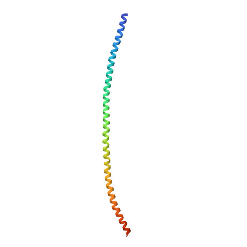
    The DNA repair factor CtIP has a critical function in double-strand break (DSB) repair by homologous recombination, promoting the assembly of the repair apparatus at DNA ends and participating in DNA-end resection. However, the molecular mechanisms of CtIP function in DSB repair remain unclear. Here, we present an atomic model for the three-dimensional architecture of human CtIP, derived from a multi-disciplinary approach that includes X-ray crystallography, small-angle X-ray scattering (SAXS) and diffracted X-ray tracking (DXT). Our data show that CtIP adopts an extended dimer-of-dimers structure, in agreement with a role in bridging distant sites on chromosomal DNA during the recombinational repair. The zinc-binding motif in the CtIP N-terminus alters dynamically the coiled-coil structure, with functional implications for the long-range interactions of CtIP with DNA. Our results provide a structural basis for the three-dimensional arrangement of chains in the CtIP tetramer, a key aspect of CtIP function in DNA DSB repair.


  • Organizational Affiliation
    • Department of Biochemistry, University of Cambridge, Cambridge CB2 1GA, UK.

Macromolecule Content 

  • Total Structure Weight: 30.55 kDa 
  • Atom Count: 1,911 
  • Modeled Residue Count: 220 
  • Deposited Residue Count: 254 
  • Unique protein chains: 1

Macromolecules

Find similar proteins by:|  3D Structure
Entity ID: 1
MoleculeChains  Sequence LengthOrganismDetailsImage
DNA endonuclease RBBP8,CtIP/RBBP8
A, B
127Homo sapiensMutation(s): 2 
Gene Names: RBBP8CTIP
EC: 3.1
UniProt & NIH Common Fund Data Resources
Find proteins for Q99708 (Homo sapiens)
Explore Q99708 
Go to UniProtKB:  Q99708
PHAROS:  Q99708
GTEx:  ENSG00000101773 
Entity Groups
Sequence Clusters30% Identity50% Identity70% Identity90% Identity95% Identity100% Identity
UniProt GroupQ99708
Sequence Annotations
Expand
Reference Sequence

Experimental Data & Validation

Experimental Data

  • Method: X-RAY DIFFRACTION
  • Resolution: 2.80 Å
  • R-Value Free:  0.253 (Depositor), 0.256 (DCC) 
  • R-Value Work:  0.224 (Depositor), 0.228 (DCC) 
Space Group: P 32
Unit Cell:
Length ( Å )Angle ( ˚ )
a = 86.598α = 90
b = 86.598β = 90
c = 42.601γ = 120
Software Package:
Software NamePurpose
PHENIXrefinement
XDSdata reduction
Aimlessdata scaling
PHASERphasing

Structure Validation

View Full Validation Report


Entry History 

& Funding Information

Deposition Data


Funding OrganizationLocationGrant Number
Wellcome TrustUnited Kingdom104641/Z/14/Z

Revision History  (Full details and data files)

  • Version 1.0: 2021-06-30
    Type: Initial release
  • Version 1.1: 2024-05-01
    Changes: Data collection, Database references, Refinement description