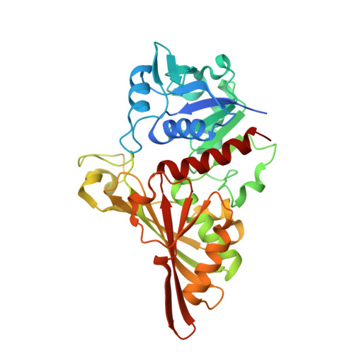
7C5P

Crystal Structure Glyceraldehyde-3-phosphate-dehydrogenase1 from Escherichia coli complexed with G3P at 2.35 Angstrom resolution.


Experimental Data Snapshot

  • Method: X-RAY DIFFRACTION
  • Resolution: 2.35 Å
  • R-Value Free: 0.210 
  • R-Value Work: 0.155 
  • R-Value Observed: 0.158 

Starting Model: experimental
View more details

wwPDB Validation   3D Report Full Report


Ligand Structure Quality Assessment 


This is version 1.3 of the entry. See complete history


Literature

Novel Structures of Type 1 Glyceraldehyde-3-phosphate Dehydrogenase from Escherichia coli Provide New Insights into the Mechanism of Generation of 1,3-Bisphosphoglyceric Acid.

Zhang, L.Liu, M.Bao, L.Bostrom, K.I.Yao, Y.Li, J.Gu, S.Ji, C.

(2021) Biomolecules 11

  • DOI: https://doi.org/10.3390/biom11111565
  • Primary Citation of Related Structures:  
    7C5G, 7C5H, 7C5I, 7C5J, 7C5K, 7C5L, 7C5M, 7C5N, 7C5O, 7C5P, 7C5Q, 7C5R, 7C7K

  • PubMed Abstract: 

    Glyceraldehyde-3-phosphate dehydrogenase (GAPDH) is a highly conserved enzyme involved in the ubiquitous process of glycolysis and presents a loop (residues 208-215 of Escherichia coli GAPDH) in two alternative conformations (I and II). It is uncertain what triggers this loop rearrangement, as well as which is the precise site from which phosphate attacks the thioacyl intermediate precursor of 1,3-bisphosphoglycerate (BPG). To clarify these uncertainties, we determined the crystal structures of complexes of wild-type GAPDH (WT) with NAD and phosphate or G3P, and of essentially inactive GAPDH mutants (C150S, H177A), trapping crystal structures for the thioacyl intermediate or for ternary complexes with NAD and either phosphate, BPG, or G3P. Analysis of these structures reported here lead us to propose that phosphate is located in the "new Pi site" attacks the thioester bond of the thioacyl intermediate to generate 1,3-bisphosphoglyceric acid (BPG). In the structure of the thioacyl intermediate, the mobile loop is in conformation II in subunits O, P, and R, while both conformations coexist in subunit Q. Moreover, only the Q subunit hosts bound NADH. In the R subunit, only the pyrophosphate part of NADH is well defined, and NADH is totally absent from the O and P subunits. Thus, the change in loop conformation appears to occur after NADH is produced, before NADH is released. In addition, two new D-glyceraldehyde-3-phosphate (G3P) binding forms are observed in WT.NAD.G3P and C150A+H177A.NAD.G3P. In summary, this paper improves our understanding of the GAPDH catalytic mechanism, particularly regarding BPG formation.


  • Organizational Affiliation

    State Key Laboratory of Genetic Engineering, School of Life Sciences, Fudan University, Shanghai 200438, China.


Macromolecules
Find similar proteins by:  (by identity cutoff)  |  3D Structure
Entity ID: 1
MoleculeChains Sequence LengthOrganismDetailsImage
Glyceraldehyde-3-phosphate dehydrogenaseA [auth O],
B [auth P],
C [auth Q],
D [auth R]
352Escherichia coli BL21(DE3)Mutation(s): 0 
Gene Names: ECBD_2224
EC: 1.2.1
UniProt
Find proteins for A0A140NCK4 (Escherichia coli (strain B / BL21-DE3))
Explore A0A140NCK4 
Go to UniProtKB:  A0A140NCK4
Entity Groups  
Sequence Clusters30% Identity50% Identity70% Identity90% Identity95% Identity100% Identity
UniProt GroupA0A140NCK4
Sequence Annotations
Expand
  • Reference Sequence
Small Molecules
Ligands 4 Unique
IDChains Name / Formula / InChI Key2D Diagram3D Interactions
NAD (Subject of Investigation/LOI)
Query on NAD

Download Ideal Coordinates CCD File 
E [auth O],
K [auth P],
P [auth Q],
T [auth R]
NICOTINAMIDE-ADENINE-DINUCLEOTIDE
C21 H27 N7 O14 P2
BAWFJGJZGIEFAR-NNYOXOHSSA-N
G3H (Subject of Investigation/LOI)
Query on G3H

Download Ideal Coordinates CCD File 
F [auth O],
L [auth P],
Q,
U [auth R]
GLYCERALDEHYDE-3-PHOSPHATE
C3 H7 O6 P
LXJXRIRHZLFYRP-VKHMYHEASA-N
PO4 (Subject of Investigation/LOI)
Query on PO4

Download Ideal Coordinates CCD File 
G [auth O],
H [auth O],
M [auth P],
R [auth Q],
V [auth R]
PHOSPHATE ION
O4 P
NBIIXXVUZAFLBC-UHFFFAOYSA-K
CL (Subject of Investigation/LOI)
Query on CL

Download Ideal Coordinates CCD File 
I [auth O],
J [auth O],
N [auth P],
O [auth P],
S [auth Q]
CHLORIDE ION
Cl
VEXZGXHMUGYJMC-UHFFFAOYSA-M
Experimental Data & Validation

Experimental Data

  • Method: X-RAY DIFFRACTION
  • Resolution: 2.35 Å
  • R-Value Free: 0.210 
  • R-Value Work: 0.155 
  • R-Value Observed: 0.158 
  • Space Group: P 41 21 2
Unit Cell:
Length ( Å )Angle ( ˚ )
a = 90.339α = 90
b = 90.339β = 90
c = 341.547γ = 90
Software Package:
Software NamePurpose
REFMACrefinement
HKL-3000data reduction
HKL-3000data scaling
MLPHAREphasing

Structure Validation

View Full Validation Report


Ligand Structure Quality Assessment 


Entry History & Funding Information

Deposition Data


Funding OrganizationLocationGrant Number
National Key Research and Development Program of ChinaChina2016YFA0500600
Science and Technology Research Program of ShanghaiChina19DZ2282100

Revision History  (Full details and data files)

  • Version 1.0: 2021-05-26
    Type: Initial release
  • Version 1.1: 2021-11-03
    Changes: Database references
  • Version 1.2: 2022-02-16
    Changes: Database references
  • Version 1.3: 2023-11-29
    Changes: Data collection, Refinement description