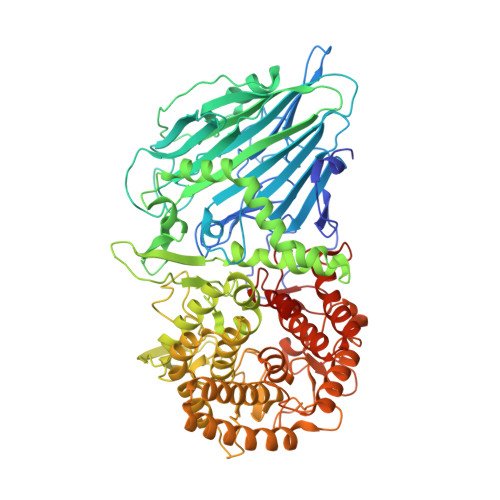
7DKU

Crystal structure of TxGH116 E441A nucleophile mutant from Thermoanaerobacterium xylanolyticum with cellobiose


Experimental Data Snapshot

  • Method: X-RAY DIFFRACTION
  • Resolution: 1.60 Å
  • R-Value Free: 0.180 
  • R-Value Work: 0.156 
  • R-Value Observed: 0.157 

Starting Model: experimental
View more details

wwPDB Validation   3D Report Full Report


Ligand Structure Quality Assessment 


This is version 1.1 of the entry. See complete history


Literature

Structural basis for transglycosylation in glycoside hydrolase family GH116 glycosynthases.

Pengthaisong, S.Hua, Y.Ketudat Cairns, J.R.

(2021) Arch Biochem Biophys 706: 108924-108924

  • DOI: https://doi.org/10.1016/j.abb.2021.108924
  • Primary Citation of Related Structures:  
    7DKS, 7DKT, 7DKU, 7DKV, 7DKW, 7DKX, 7DKY

  • PubMed Abstract: 

    Glycosynthases are glycoside hydrolase mutants that can synthesize oligosaccharides or glycosides from an inverted donor without hydrolysis of the products. Although glycosynthases have been characterized from a variety of glycoside hydrolase (GH) families, family GH116 glycosynthases have yet to be reported. We produced the Thermoanaerobacterium xylanolyticum TxGH116 nucleophile mutants E441D, E441G, E441Q and E441S and compared their glycosynthase activities to the previously generated E441A mutant. The TxGH116 E441G and E441S mutants exhibited highest glycosynthase activity to transfer glucose from α-fluoroglucoside (α-GlcF) to cellobiose acceptor, while E441D had low but significant activity as well. The E441G, E441S and E441A variants showed broad specificity for α-glycosyl fluoride donors and p-nitrophenyl glycoside acceptors. The structure of the TxGH116 E441A mutant with α-GlcF provided the donor substrate complex, while soaking of the TxGH116 E441G mutant with α-GlcF resulted in cellooligosaccharides extending from the +1 subsite out of the active site, with glycerol in the -1 subsite. Soaking of E441A or E441G with cellobiose or cellotriose gave similar acceptor substrate complexes with the nonreducing glucosyl residue in the +1 subsite. Combining structures with the ligands from the TxGH116 E441A with α-GlcF crystals with that of E441A or E441G with cellobiose provides a plausible structure of the catalytic ternary complex, which places the nonreducing glucosyl residue O4 2.5 Å from the anomeric carbon of α-GlcF, thereby explaining its apparent preference for production of β-1,4-linked oligosaccharides. This functional and structural characterization provides the background for development of GH116 glycosynthases for synthesis of oligosaccharides and glycosides of interest.


  • Organizational Affiliation

    School of Chemistry, Institute of Science, Suranaree University of Technology, Nakhon Ratchasima, 30000, Thailand; Center for Biomolecular Structure, Function and Application, Suranaree University of Technology, Nakhon Ratchasima, 30000, Thailand.


Macromolecules
Find similar proteins by:  (by identity cutoff)  |  3D Structure
Entity ID: 1
MoleculeChains Sequence LengthOrganismDetailsImage
beta-glucosidase799Thermoanaerobacterium xylanolyticum LX-11Mutation(s): 1 
Gene Names: Thexy_2211
EC: 3.2.1.21 (PDB Primary Data), 3.2.1.45 (UniProt)
UniProt
Find proteins for F6BL85 (Thermoanaerobacterium xylanolyticum (strain ATCC 49914 / DSM 7097 / LX-11))
Explore F6BL85 
Go to UniProtKB:  F6BL85
Entity Groups  
Sequence Clusters30% Identity50% Identity70% Identity90% Identity95% Identity100% Identity
UniProt GroupF6BL85
Sequence Annotations
Expand
  • Reference Sequence
Oligosaccharides

Help

Entity ID: 2
MoleculeChains Length2D Diagram Glycosylation3D Interactions
beta-D-glucopyranose-(1-4)-beta-D-glucopyranose
B
2N/A
Glycosylation Resources
GlyTouCan:  G84824ZO
GlyCosmos:  G84824ZO
Small Molecules
Ligands 2 Unique
IDChains Name / Formula / InChI Key2D Diagram3D Interactions
GOL (Subject of Investigation/LOI)
Query on GOL

Download Ideal Coordinates CCD File 
C [auth A]
D [auth A]
E [auth A]
F [auth A]
G [auth A]
C [auth A],
D [auth A],
E [auth A],
F [auth A],
G [auth A],
H [auth A],
I [auth A],
J [auth A],
K [auth A],
L [auth A],
M [auth A],
N [auth A],
O [auth A],
P [auth A]
GLYCEROL
C3 H8 O3
PEDCQBHIVMGVHV-UHFFFAOYSA-N
CA (Subject of Investigation/LOI)
Query on CA

Download Ideal Coordinates CCD File 
Q [auth A]CALCIUM ION
Ca
BHPQYMZQTOCNFJ-UHFFFAOYSA-N
Experimental Data & Validation

Experimental Data

  • Method: X-RAY DIFFRACTION
  • Resolution: 1.60 Å
  • R-Value Free: 0.180 
  • R-Value Work: 0.156 
  • R-Value Observed: 0.157 
  • Space Group: P 21 21 2
Unit Cell:
Length ( Å )Angle ( ˚ )
a = 177.259α = 90
b = 54.459β = 90
c = 83.154γ = 90
Software Package:
Software NamePurpose
REFMACrefinement
PDB_EXTRACTdata extraction
Cootmodel building
HKL-2000data scaling
MOLREPphasing
HKL-2000data reduction

Structure Validation

View Full Validation Report


Ligand Structure Quality Assessment 


Entry History & Funding Information

Deposition Data


Funding OrganizationLocationGrant Number
Other privateThailandThailand Research Fund
Other privateThailandSuranaree University of Technology

Revision History  (Full details and data files)

  • Version 1.0: 2021-06-23
    Type: Initial release
  • Version 1.1: 2023-11-29
    Changes: Data collection, Database references, Refinement description