Mutational Biosynthesis of Hitachimycin Analogs Controlled by the beta-Amino Acid-Selective Adenylation Enzyme HitB.
Kudo, F., Takahashi, S., Miyanaga, A., Nakazawa, Y., Nishino, K., Hayakawa, Y., Kawamura, K., Ishikawa, F., Tanabe, G., Iwai, N., Nagumo, Y., Usui, T., Eguchi, T.(2021) ACS Chem Biol 16: 539-547
- PubMed: 33625847
- DOI: https://doi.org/10.1021/acschembio.1c00003
- Primary Citation of Related Structures:
7DQ5, 7DQ6 - PubMed Abstract:
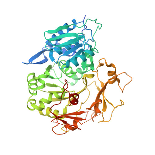
Hitachimycin is a macrolactam antibiotic with an ( S )-β-phenylalanine (β-Phe) at the starter position of its polyketide skeleton. ( S )-β-Phe is formed from l-α-phenylalanine by the phenylananine-2,3-aminomutase HitA in the hitachimycin biosynthetic pathway. In this study, we produced new hitachimycin analogs via mutasynthesis by feeding various ( S )-β-Phe analogs to a Δ hitA strain. We obtained six hitachimycin analogs with F at the ortho , meta , or para position and Cl, Br, or a CH 3 group at the meta position of the phenyl moiety, as well as two hitachimycin analogs with thienyl substitutions. Furthermore, we carried out a biochemical and structural analysis of HitB, a β-amino acid-selective adenylation enzyme that introduces ( S )-β-Phe into the hitachimycin biosynthetic pathway. The K M values of the incorporated ( S )-β-Phe analogs and natural ( S )-β-Phe were similar. However, the K M values of unincorporated ( S )-β-Phe analogs with Br and a CH 3 group at the ortho or para position of the phenyl moiety were high, indicating that HitB functions as a gatekeeper to select macrolactam starter units during mutasynthesis. The crystal structure of HitB in complex with ( S )-β-3-Br-phenylalanine sulfamoyladenosine (β- m -Br-Phe-SA) revealed that the bulky meta- Br group is accommodated by the conformational flexibility around Phe328, whose side chain is close to the meta position. The aromatic group of β- m -Br-Phe-SA is surrounded by hydrophobic and aromatic residues, which appears to confer the conformational flexibility that enables HitB to accommodate the meta -substituted ( S )-β-Phe. The new hitachimycin analogs exhibited different levels of biological activity in HeLa cells and multidrug-sensitive budding yeast, suggesting that they may target different molecules.
- Department of Chemistry, Tokyo Institute of Technology, 2-12-1 Meguro-ku, O-okayama, Tokyo 152-8551, Japan.
Organizational Affiliation:


