7N9G

Crystal structure of the Abl 1b Kinase domain in complex with Dasatinib and Imatinib


Experimental Data Snapshot

  • Method: X-RAY DIFFRACTION
  • Resolution: 2.20 Å
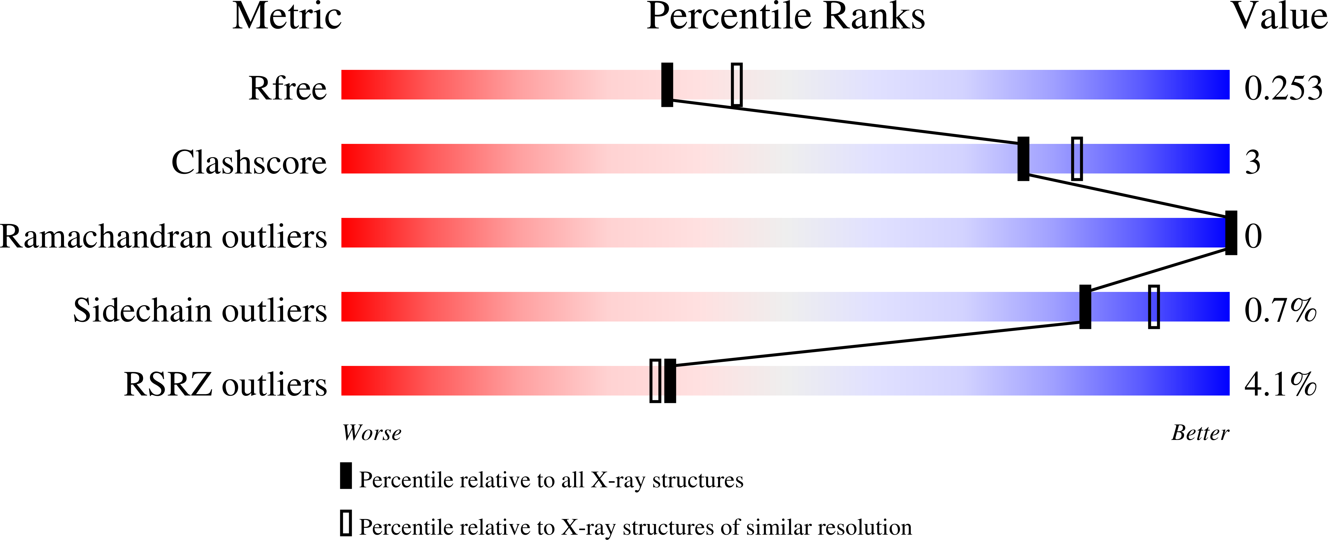
  • R-Value Free: 0.251 
  • R-Value Work: 0.217 

Starting Model: experimental
View more details

wwPDB Validation   3D Report Full Report


Ligand Structure Quality Assessment 


This is version 1.1 of the entry. See complete history


Literature

Imatinib can act as an Allosteric Activator of Abl Kinase.

Xie, T.Saleh, T.Rossi, P.Miller, D.Kalodimos, C.G.

(2022) J Mol Biol 434: 167349-167349

  • DOI: https://doi.org/10.1016/j.jmb.2021.167349
  • Primary Citation of Related Structures:  
    7N9G

  • PubMed Abstract: 

    Imatinib is an ATP-competitive inhibitor of Bcr-Abl kinase and the first drug approved for chronic myelogenous leukemia (CML) treatment. Here we show that imatinib binds to a secondary, allosteric site located in the myristoyl pocket of Abl to function as an activator of the kinase activity. Abl transitions between an assembled, inhibited state and an extended, activated state. The equilibrium is regulated by the conformation of the αΙ helix, which is located nearby the allosteric pocket. Imatinib binding to the allosteric pocket elicits an αΙ helix conformation that is not compatible with the assembled state, thereby promoting the extended state and stimulating the kinase activity. Although in wild-type Abl the catalytic pocket has a much higher affinity for imatinib than the allosteric pocket does, the two binding affinities are comparable in Abl variants carrying imatinib-resistant mutations in the catalytic site. A previously isolated imatinib-resistant mutation in patients appears to be mediating its function by increasing the affinity of imatinib for the allosteric pocket, providing a hitherto unknown mechanism of drug resistance. Our results highlight the benefit of combining imatinib with allosteric inhibitors to maximize their inhibitory effect on Bcr-Abl.


  • Organizational Affiliation

    Department of Structural Biology, St. Jude Children's Research Hospital, Memphis, TN 38105, United States.


Macromolecules
Find similar proteins by:  (by identity cutoff)  |  3D Structure
Entity ID: 1
MoleculeChains Sequence LengthOrganismDetailsImage
Tyrosine-protein kinase ABL1
A, B, C
271Homo sapiensMutation(s): 0 
Gene Names: ABL1ABLJTK7
EC: 2.7.10.2
UniProt & NIH Common Fund Data Resources
Find proteins for P00519 (Homo sapiens)
Explore P00519 
Go to UniProtKB:  P00519
PHAROS:  P00519
GTEx:  ENSG00000097007 
Entity Groups  
Sequence Clusters30% Identity50% Identity70% Identity90% Identity95% Identity100% Identity
UniProt GroupP00519
Sequence Annotations
Expand
  • Reference Sequence
Small Molecules
Ligands 3 Unique
IDChains Name / Formula / InChI Key2D Diagram3D Interactions
STI (Subject of Investigation/LOI)
Query on STI

Download Ideal Coordinates CCD File 
D [auth A],
G [auth B],
I [auth C]
4-(4-METHYL-PIPERAZIN-1-YLMETHYL)-N-[4-METHYL-3-(4-PYRIDIN-3-YL-PYRIMIDIN-2-YLAMINO)-PHENYL]-BENZAMIDE
C29 H31 N7 O
KTUFNOKKBVMGRW-UHFFFAOYSA-N
1N1 (Subject of Investigation/LOI)
Query on 1N1

Download Ideal Coordinates CCD File 
E [auth A],
H [auth B],
J [auth C]
N-(2-CHLORO-6-METHYLPHENYL)-2-({6-[4-(2-HYDROXYETHYL)PIPERAZIN-1-YL]-2-METHYLPYRIMIDIN-4-YL}AMINO)-1,3-THIAZOLE-5-CARBOXAMIDE
C22 H26 Cl N7 O2 S
ZBNZXTGUTAYRHI-UHFFFAOYSA-N
PO4
Query on PO4

Download Ideal Coordinates CCD File 
F [auth A]PHOSPHATE ION
O4 P
NBIIXXVUZAFLBC-UHFFFAOYSA-K
Binding Affinity Annotations 
IDSourceBinding Affinity
STI BindingDB:  7N9G Ki: min: 13, max: 7000 (nM) from 5 assay(s)
Kd: min: 1, max: 1.00e+4 (nM) from 35 assay(s)
IC50: min: 1.1, max: 1.00e+4 (nM) from 68 assay(s)
EC50: min: 34, max: 1000 (nM) from 4 assay(s)
1N1 BindingDB:  7N9G Kd: min: 0.02, max: 120 (nM) from 27 assay(s)
IC50: min: 0.14, max: 1140 (nM) from 24 assay(s)
EC50: 0.04 (nM) from 1 assay(s)
Experimental Data & Validation

Experimental Data

  • Method: X-RAY DIFFRACTION
  • Resolution: 2.20 Å
  • R-Value Free: 0.251 
  • R-Value Work: 0.217 
  • Space Group: I 2 2 2
Unit Cell:
Length ( Å )Angle ( ˚ )
a = 69.933α = 90
b = 166.879β = 90
c = 186.817γ = 90
Software Package:
Software NamePurpose
REFMACrefinement
XDSdata reduction
Aimlessdata scaling
PHASERphasing

Structure Validation

View Full Validation Report


Ligand Structure Quality Assessment 


Entry History & Funding Information

Deposition Data


Funding OrganizationLocationGrant Number
National Institutes of Health/Office of the DirectorUnited States5R35GM122462-05

Revision History  (Full details and data files)

  • Version 1.0: 2022-04-27
    Type: Initial release
  • Version 1.1: 2023-10-18
    Changes: Data collection, Refinement description