7P2I | pdb_00007p2i

Dimethylated fusion protein of RSL and nucleoporin peptide (Nup) in complex with cucurbit[7]uril, F432 cage assembly


Experimental Data Snapshot

  • Method: X-RAY DIFFRACTION
  • Resolution: 1.49 Å
  • R-Value Free: 
    0.178 (Depositor), 0.185 (DCC) 
  • R-Value Work: 
    0.162 (Depositor), 0.162 (DCC) 
  • R-Value Observed: 
    0.162 (Depositor) 

Starting Model: experimental
View more details

wwPDB Validation 3D Report Full Report

Validation slider image for 7P2I

Ligand Structure Quality Assessment 


This is version 1.2 of the entry. See complete history

Literature

Segregated Protein-Cucurbit[7]uril Crystalline Architectures via Modulatory Peptide Tectons.

Ramberg, K.O.Guagnini, F.Engilberge, S.Wronska, M.A.Rennie, M.L.Perez, J.Crowley, P.B.

(2021) Chemistry 27: 14619-14627

  • DOI: https://doi.org/10.1002/chem.202103025
  • Primary Citation Related Structures: 
    6S99, 7P2H, 7P2I, 7P2J

  • PubMed Abstract: 

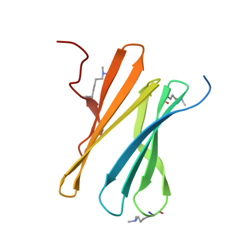
    One approach to protein assembly involves water-soluble supramolecular receptors that act like glues. Bionanoarchitectures directed by these scaffolds are often system-specific, with few studies investigating their customization. Herein, the modulation of cucurbituril-mediated protein assemblies through the inclusion of peptide tectons is described. Three peptides of varying length and structural order were N-terminally appended to RSL, a β-propeller building block. Each fusion protein was incorporated into crystalline architectures mediated by cucurbit[7]uril (Q7). A trimeric coiled-coil served as a spacer within a Q7-directed sheet assembly of RSL, giving rise to a layered material of varying porosity. Within the spacer layers, the coiled-coils were dynamic. This result prompted consideration of intrinsically disordered peptides (IDPs) as modulatory tectons. Similar to the coiled-coil, a mussel adhesion peptide (Mefp) also acted as a spacer between protein-Q7 sheets. In contrast, the fusion of a nucleoporin peptide (Nup) to RSL did not recapitulate the sheet assembly. Instead, a Q7-directed cage was adopted, within which disordered Nup peptides were partially "captured" by Q7 receptors. IDP capture occurred by macrocycle recognition of an intrapeptide Phe-Gly motif in which the benzyl group was encapsulated by Q7. The modularity of these protein-cucurbituril architectures adds a new dimension to macrocycle-mediated protein assembly. Segregated protein crystals, with alternating layers of high and low porosity, could provide a basis for new types of materials.


  • Organizational Affiliation
    • School of Chemistry, National University of Ireland Galway, University Road, Galway, H91 TK33, Ireland.

Macromolecule Content 

  • Total Structure Weight: 36.62 kDa 
  • Atom Count: 2,809 
  • Modeled Residue Count: 271 
  • Deposited Residue Count: 306 
  • Unique protein chains: 1

Macromolecules

Find similar proteins by:|  3D Structure
Entity ID: 1
MoleculeChains  Sequence LengthOrganismDetailsImage
Fucose-binding lectin protein
A, B, C
102Ralstonia solanacearumMutation(s): 0 
Gene Names: E7Z57_08365HXP36_18875RSP795_21825RSP822_19650RUN39_v1_50103
UniProt
Find proteins for A0A0S4TLR1 (Ralstonia solanacearum)
Explore A0A0S4TLR1 
Go to UniProtKB:  A0A0S4TLR1
Entity Groups
Sequence Clusters30% Identity50% Identity70% Identity90% Identity95% Identity100% Identity
UniProt GroupA0A0S4TLR1
Sequence Annotations
Expand
Reference Sequence

Small Molecules

Ligands 2 Unique
IDChains Name / Formula / InChI Key2D Diagram3D Interactions
QQ7
(Subject of Investigation/LOI)

Query on QQ7


Download:Ideal Coordinates CCD File
F [auth A],
K [auth C]
cucurbit[7]uril
C42 H42 N28 O14
ZDOBFUIMGBWEAB-XGFHMVPTSA-N
MFU

Query on MFU


Download:Ideal Coordinates CCD File
D [auth A]
E [auth A]
G [auth B]
H [auth B]
I [auth C]
D [auth A],
E [auth A],
G [auth B],
H [auth B],
I [auth C],
J [auth C]
methyl alpha-L-fucopyranoside
C7 H14 O5
OHWCAVRRXKJCRB-CXNFULCWSA-N
Modified Residues  1 Unique
IDChains TypeFormula2D DiagramParent
MLY
Query on MLY
A, B, C
L-PEPTIDE LINKINGC8 H18 N2 O2LYS

Experimental Data & Validation

Experimental Data

  • Method: X-RAY DIFFRACTION
  • Resolution: 1.49 Å
  • R-Value Free:  0.178 (Depositor), 0.185 (DCC) 
  • R-Value Work:  0.162 (Depositor), 0.162 (DCC) 
  • R-Value Observed: 0.162 (Depositor) 
Space Group: F 4 3 2
Unit Cell:
Length ( Å )Angle ( ˚ )
a = 200.713α = 90
b = 200.713β = 90
c = 200.713γ = 90
Software Package:
Software NamePurpose
BUSTERrefinement
PDB_EXTRACTdata extraction
XDSdata reduction
Aimlessdata scaling
BUSTERphasing

Structure Validation

View Full Validation Report


Ligand Structure Quality Assessment 


Entry History 

& Funding Information

Deposition Data


Funding OrganizationLocationGrant Number
Science Foundation IrelandIreland13/CDA/2168
Science Foundation IrelandIrelandSSPC-PharM5 12/RC/2275_P2
Irish Research CouncilIrelandGOIPD/2019/513

Revision History  (Full details and data files)

  • Version 1.0: 2021-09-08
    Type: Initial release
  • Version 1.1: 2021-11-03
    Changes: Database references
  • Version 1.2: 2024-01-31
    Changes: Data collection, Refinement description