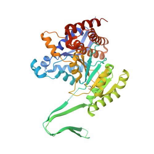
7PJN

Crystal Structure of Ivosidenib-resistant IDH1 variant R132C S280F in complex with NADPH and inhibitor DS-1001B


Experimental Data Snapshot

  • Method: X-RAY DIFFRACTION
  • Resolution: 2.45 Å
  • R-Value Free: 0.228 
  • R-Value Work: 0.186 
  • R-Value Observed: 0.188 

Starting Model: experimental
View more details

wwPDB Validation   3D Report Full Report


This is version 1.3 of the entry. See complete history


Literature

Resistance to the isocitrate dehydrogenase 1 mutant inhibitor ivosidenib can be overcome by alternative dimer-interface binding inhibitors.

Reinbold, R.Hvinden, I.C.Rabe, P.Herold, R.A.Finch, A.Wood, J.Morgan, M.Staudt, M.Clifton, I.J.Armstrong, F.A.McCullagh, J.S.O.Redmond, J.Bardella, C.Abboud, M.I.Schofield, C.J.

(2022) Nat Commun 13: 4785-4785

  • DOI: https://doi.org/10.1038/s41467-022-32436-4
  • Primary Citation of Related Structures:  
    7PJM, 7PJN

  • PubMed Abstract: 

    Ivosidenib, an inhibitor of isocitrate dehydrogenase 1 (IDH1) R132C and R132H variants, is approved for the treatment of acute myeloid leukaemia (AML). Resistance to ivosidenib due to a second site mutation of IDH1 R132C, leading to IDH1 R132C/S280F, has emerged. We describe biochemical, crystallographic, and cellular studies on the IDH1 R132C/S280F and R132H/S280F variants that inform on the mechanism of second-site resistance, which involves both modulation of inhibitor binding at the IDH1 dimer-interface and alteration of kinetic properties, which enable more efficient 2-HG production relative to IDH1 R132C and IDH1 R132H. Importantly, the biochemical and cellular results demonstrate that it should be possible to overcome S280F mediated resistance in AML patients by using alternative inhibitors, including some presently in phase 2 clinical trials.


  • Organizational Affiliation

    Chemistry Research Laboratory, Department of Chemistry and the Ineos Oxford Institute for Antimicrobial Research, University of Oxford, 12 Mansfield, Oxford, OX1 3TA, UK.


Macromolecules
Find similar proteins by:  (by identity cutoff)  |  3D Structure
Entity ID: 1
MoleculeChains Sequence LengthOrganismDetailsImage
Isocitrate dehydrogenase [NADP] cytoplasmic
A, C, D
422Homo sapiensMutation(s): 2 
Gene Names: IDH1PICD
EC: 1.1.1.42
UniProt & NIH Common Fund Data Resources
Find proteins for O75874 (Homo sapiens)
Explore O75874 
Go to UniProtKB:  O75874
PHAROS:  O75874
GTEx:  ENSG00000138413 
Entity Groups  
Sequence Clusters30% Identity50% Identity70% Identity90% Identity95% Identity100% Identity
UniProt GroupO75874
Sequence Annotations
Expand
  • Reference Sequence
Find similar proteins by:  (by identity cutoff)  |  3D Structure
Entity ID: 2
MoleculeChains Sequence LengthOrganismDetailsImage
Isocitrate dehydrogenase [NADP] cytoplasmic422Homo sapiensMutation(s): 2 
Gene Names: IDH1PICD
EC: 1.1.1.42
UniProt & NIH Common Fund Data Resources
Find proteins for O75874 (Homo sapiens)
Explore O75874 
Go to UniProtKB:  O75874
PHAROS:  O75874
GTEx:  ENSG00000138413 
Entity Groups  
Sequence Clusters30% Identity50% Identity70% Identity90% Identity95% Identity100% Identity
UniProt GroupO75874
Sequence Annotations
Expand
  • Reference Sequence
Small Molecules
Ligands 4 Unique
IDChains Name / Formula / InChI Key2D Diagram3D Interactions
NDP (Subject of Investigation/LOI)
Query on NDP

Download Ideal Coordinates CCD File 
H [auth A],
M [auth B],
S [auth C],
W [auth D]
NADPH DIHYDRO-NICOTINAMIDE-ADENINE-DINUCLEOTIDE PHOSPHATE
C21 H30 N7 O17 P3
ACFIXJIJDZMPPO-NNYOXOHSSA-N
7SU (Subject of Investigation/LOI)
Query on 7SU

Download Ideal Coordinates CCD File 
E [auth A],
J [auth B],
O [auth C],
T [auth D]
(E)-3-(1-(5-(2-fluoropropan-2-yl)-3-(2,4,6-trichlorophenyl)isoxazole-4-carbonyl)-3-methyl-1H-indol-4-yl)acrylic acid
C25 H18 Cl3 F N2 O4
BOOMBLZEOHXPPX-BQYQJAHWSA-N
CIT
Query on CIT

Download Ideal Coordinates CCD File 
F [auth A],
I [auth A],
L [auth B],
Q [auth C],
V [auth D]
CITRIC ACID
C6 H8 O7
KRKNYBCHXYNGOX-UHFFFAOYSA-N
GOL
Query on GOL

Download Ideal Coordinates CCD File 
G [auth A]
K [auth B]
N [auth B]
P [auth C]
R [auth C]
G [auth A],
K [auth B],
N [auth B],
P [auth C],
R [auth C],
U [auth D]
GLYCEROL
C3 H8 O3
PEDCQBHIVMGVHV-UHFFFAOYSA-N
Modified Residues  1 Unique
IDChains TypeFormula2D DiagramParent
CSD
Query on CSD
B
L-PEPTIDE LINKINGC3 H7 N O4 SCYS
Experimental Data & Validation

Experimental Data

  • Method: X-RAY DIFFRACTION
  • Resolution: 2.45 Å
  • R-Value Free: 0.228 
  • R-Value Work: 0.186 
  • R-Value Observed: 0.188 
  • Space Group: P 21 21 21
Unit Cell:
Length ( Å )Angle ( ˚ )
a = 80.18α = 90
b = 153.05β = 90
c = 164.01γ = 90
Software Package:
Software NamePurpose
PHENIXrefinement
Aimlessdata scaling
PDB_EXTRACTdata extraction
xia2data reduction
PHASERphasing

Structure Validation

View Full Validation Report


Entry History & Funding Information

Deposition Data


Funding OrganizationLocationGrant Number
Wellcome TrustUnited Kingdom106244/Z/14/Z
Biotechnology and Biological Sciences Research Council (BBSRC)United KingdomBB/R506655/1

Revision History  (Full details and data files)

  • Version 1.0: 2022-07-13
    Type: Initial release
  • Version 1.1: 2022-10-19
    Changes: Database references
  • Version 1.2: 2024-01-31
    Changes: Data collection, Refinement description
  • Version 1.3: 2024-10-09
    Changes: Structure summary