7R0F | pdb_00007r0f

Crystal structure of a flavodiiron protein D52K mutant in the oxidized state from Escherichia coli


Experimental Data Snapshot

  • Method: X-RAY DIFFRACTION
  • Resolution: 1.98 Å
  • R-Value Free: 
    0.205 (Depositor), 0.216 (DCC) 
  • R-Value Work: 
    0.205 (Depositor), 0.230 (DCC) 
  • R-Value Observed: 
    0.205 (Depositor) 

Starting Model: other
View more details

wwPDB Validation 3D Report Full Report

Validation slider image for 7R0F

Ligand Structure Quality Assessment 


This is version 1.2 of the entry. See complete history

Literature

Structures of variants of Escherichia coli flavodiiron-type nitric oxide reductase reveal changes in the di-iron site.

Borges, P.T.Folgosa, F.Martins, M.C.Gotthard, G.van der Linden, P.Carpentier, P.Teixeira, M.Frazao, C.Romao, C.V.

(2026) Acta Crystallogr D Struct Biol 82: 457-470

  • DOI: https://doi.org/10.1107/S2059798326002214
  • Primary Citation Related Structures: 
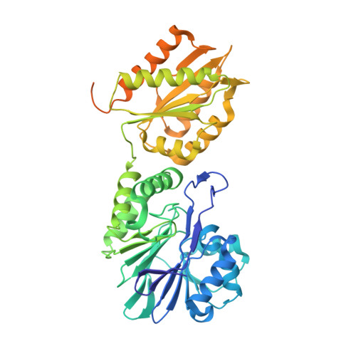
    7R0F, 7R1H, 7R2S

  • PubMed Abstract: 

    Flavodiiron proteins (FDPs) are NO and/or O 2 reductases which contain a di-iron catalytic center. Interestingly, they exhibit different selectivities towards each one of these substrates, despite having the same ligands of the iron ions. Escherichia coli FDP is a selective NO reductase that protects this bacterium against nitric oxide by catalyzing two-electron reduction to the nontoxic N 2 O. Previously, based on kinetic studies, we explored the possible role of two amino acids located in the di-iron second coordination sphere, Lys53 and Tyr271, in modulation of the substrate selectivity of Entamoeba histolytica FDP, a selective O 2 reductase. In this work, we replaced the structurally equivalent residues in E. coli FDP, Asp52 and Ser262, by those present in the O 2 -selective FDP and determined their crystal structures in both oxidized and reduced states. Furthermore, the molecular-substrate tunnels were experimentally identified using krypton pressurization of the crystals. The data obtained corroborated previous molecular-dynamics calculations on this FDP. The side chains of residues in both positions 52 and 262 of E. coli FDP variants and wild type are in the vicinity of the shorter intramolecular tunnel, which is suggested to be the exit route for the reaction products N 2 O and H 2 O. The E. coli FDP S262Y variant shows photoreduction of the di-iron center and partial loss of electron density in some of its coordinating ligands after X-ray exposure, and these effects are consistent with increased radiation sensitivity. The kinetic properties of the variants towards NO and O 2 were not significantly different from the wild type, contrary to what was observed previously for E. histolytica FDP.


  • Organizational Affiliation
    • ITQB NOVA, Instituto de Tecnologia Química e Biológica António Xavier, Universidade Nova de Lisboa, Avenida da República, 2780-157 Oeiras, Portugal.

Macromolecule Content 

  • Total Structure Weight: 109.85 kDa 
  • Atom Count: 7,053 
  • Modeled Residue Count: 802 
  • Deposited Residue Count: 958 
  • Unique protein chains: 1

Macromolecules

Find similar proteins by:|  3D Structure
Entity ID: 1
MoleculeChains  Sequence LengthOrganismDetailsImage
Anaerobic nitric oxide reductase flavorubredoxin
A, B
479Escherichia coli K-12Mutation(s): 1 
Gene Names: norVflrDygaIygaJygaKb2710JW2680
UniProt
Find proteins for Q46877 (Escherichia coli (strain K12))
Explore Q46877 
Go to UniProtKB:  Q46877
Entity Groups
Sequence Clusters30% Identity50% Identity70% Identity90% Identity95% Identity100% Identity
UniProt GroupQ46877
Sequence Annotations
Expand
Reference Sequence

Small Molecules

Ligands 4 Unique
IDChains Name / Formula / InChI Key2D Diagram3D Interactions
FMN
(Subject of Investigation/LOI)

Query on FMN


Download:Ideal Coordinates CCD File
C [auth A],
H [auth B]
FLAVIN MONONUCLEOTIDE
C17 H21 N4 O9 P
FVTCRASFADXXNN-SCRDCRAPSA-N
FE

Query on FE


Download:Ideal Coordinates CCD File
D [auth A],
E [auth A],
I [auth B],
J [auth B]
FE (III) ION
Fe
VTLYFUHAOXGGBS-UHFFFAOYSA-N
OXY

Query on OXY


Download:Ideal Coordinates CCD File
G [auth A],
L [auth B]
OXYGEN MOLECULE
O2
MYMOFIZGZYHOMD-UHFFFAOYSA-N
O

Query on O


Download:Ideal Coordinates CCD File
F [auth A],
K [auth B]
OXYGEN ATOM
O
XLYOFNOQVPJJNP-UHFFFAOYSA-N

Experimental Data & Validation

Experimental Data

  • Method: X-RAY DIFFRACTION
  • Resolution: 1.98 Å
  • R-Value Free:  0.205 (Depositor), 0.216 (DCC) 
  • R-Value Work:  0.205 (Depositor), 0.230 (DCC) 
  • R-Value Observed: 0.205 (Depositor) 
Space Group: I 1 2 1
Unit Cell:
Length ( Å )Angle ( ˚ )
a = 88.958α = 90
b = 64.051β = 91.14
c = 146.48γ = 90
Software Package:
Software NamePurpose
PHENIXrefinement
XSCALEdata scaling
PDB_EXTRACTdata extraction
XDSdata reduction
PHENIXphasing

Structure Validation

View Full Validation Report


Ligand Structure Quality Assessment 


Entry History 

& Funding Information

Deposition Data


Funding OrganizationLocationGrant Number
Foundation for Science and Technology (FCT)United Kingdom--

Revision History  (Full details and data files)

  • Version 1.0: 2023-02-15
    Type: Initial release
  • Version 1.1: 2024-05-01
    Changes: Data collection, Refinement description
  • Version 1.2: 2026-06-17
    Changes: Database references, Structure summary