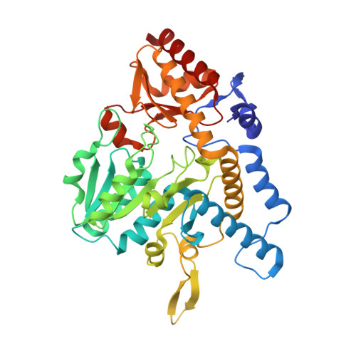
7RUJ

E. coli cysteine desulfurase SufS N99A


Experimental Data Snapshot

  • Method: X-RAY DIFFRACTION
  • Resolution: 2.50 Å
  • R-Value Free: 0.219 
  • R-Value Work: 0.193 
  • R-Value Observed: 0.194 

Starting Model: experimental
View more details

wwPDB Validation   3D Report Full Report


Ligand Structure Quality Assessment 


This is version 1.2 of the entry. See complete history


Literature

The beta-latch structural element of the SufS cysteine desulfurase mediates active site accessibility and SufE transpersulfurase positioning.

Gogar, R.K.Carroll, F.Conte, J.V.Nasef, M.Dunkle, J.A.Frantom, P.A.

(2023) J Biol Chem 299: 102966-102966

  • DOI: https://doi.org/10.1016/j.jbc.2023.102966
  • Primary Citation of Related Structures:  
    7RUJ, 7RW3

  • PubMed Abstract: 

    Under oxidative stress and iron starvation conditions, Escherichia coli uses the Suf pathway to assemble iron-sulfur clusters. The Suf pathway mobilizes sulfur via SufS, a type II cysteine desulfurase. SufS is a pyridoxal-5'-phosphate-dependent enzyme that uses cysteine to generate alanine and an active-site persulfide (C 364 -S-S - ). The SufS persulfide is protected from external oxidants/reductants and requires the transpersulfurase, SufE, to accept the persulfide to complete the SufS catalytic cycle. Recent reports on SufS identified a conserved "β-latch" structural element that includes the α 6 helix, a glycine-rich loop, a β-hairpin, and a cis-proline residue. To identify a functional role for the β-latch, we used site-directed mutagenesis to obtain the N99D and N99A SufS variants. N99 is a conserved residue that connects the α 6 helix to the backbone of the glycine-rich loop via hydrogen bonds. Our x-ray crystal structures for N99A and N99D SufS show a distorted beta-hairpin and glycine-rich loop, respectively, along with changes in the dimer geometry. The structural disruption of the N99 variants allowed the external reductant TCEP to react with the active-site C364-persulfide intermediate to complete the SufS catalytic cycle in the absence of SufE. The substitutions also appear to disrupt formation of a high-affinity, close approach SufS-SufE complex as measured with fluorescence polarization. Collectively, these findings demonstrate that the β-latch does not affect the chemistry of persulfide formation but does protect it from undesired reductants. The data also indicate the β-latch plays an unexpected role in forming a close approach SufS-SufE complex to promote persulfide transfer.


  • Organizational Affiliation

    Department of Chemistry & Biochemistry, The University of Alabama, Tuscaloosa, Alabama, USA.


Macromolecules
Find similar proteins by:  (by identity cutoff)  |  3D Structure
Entity ID: 1
MoleculeChains Sequence LengthOrganismDetailsImage
Cysteine desulfurase406Escherichia coliMutation(s): 1 
Gene Names: 
EC: 2.8.1.7 (PDB Primary Data), 4.4.1.16 (PDB Primary Data), 3.13.1 (UniProt)
UniProt
Find proteins for P77444 (Escherichia coli (strain K12))
Explore P77444 
Go to UniProtKB:  P77444
Entity Groups  
Sequence Clusters30% Identity50% Identity70% Identity90% Identity95% Identity100% Identity
UniProt GroupP77444
Sequence Annotations
Expand
  • Reference Sequence
Small Molecules
Ligands 2 Unique
IDChains Name / Formula / InChI Key2D Diagram3D Interactions
PLP (Subject of Investigation/LOI)
Query on PLP

Download Ideal Coordinates CCD File 
C [auth A]PYRIDOXAL-5'-PHOSPHATE
C8 H10 N O6 P
NGVDGCNFYWLIFO-UHFFFAOYSA-N
CL
Query on CL

Download Ideal Coordinates CCD File 
B [auth A]CHLORIDE ION
Cl
VEXZGXHMUGYJMC-UHFFFAOYSA-M
Experimental Data & Validation

Experimental Data

  • Method: X-RAY DIFFRACTION
  • Resolution: 2.50 Å
  • R-Value Free: 0.219 
  • R-Value Work: 0.193 
  • R-Value Observed: 0.194 
  • Space Group: P 43 21 2
Unit Cell:
Length ( Å )Angle ( ˚ )
a = 125.229α = 90
b = 125.229β = 90
c = 137.429γ = 90
Software Package:
Software NamePurpose
PHENIXrefinement
Aimlessdata scaling
PDB_EXTRACTdata extraction
CrysalisProdata reduction
PHENIXphasing

Structure Validation

View Full Validation Report


Ligand Structure Quality Assessment 


Entry History & Funding Information

Deposition Data


Funding OrganizationLocationGrant Number
National Institutes of Health/National Institute of General Medical Sciences (NIH/NIGMS)United States--

Revision History  (Full details and data files)

  • Version 1.0: 2023-01-25
    Type: Initial release
  • Version 1.1: 2023-08-09
    Changes: Database references
  • Version 1.2: 2023-10-25
    Changes: Data collection, Refinement description