7SCW

KRAS full length wild-type in complex with RGL1 Ras association domain


Experimental Data Snapshot

  • Method: X-RAY DIFFRACTION
  • Resolution: 1.98 Å
  • R-Value Free: 
    0.192 (Depositor), 0.190 (DCC) 
  • R-Value Work: 
    0.156 (Depositor), 0.150 (DCC) 
  • R-Value Observed: 
    0.158 (Depositor) 

Starting Model: experimental
View more details

wwPDB Validation   3D Report Full Report


Ligand Structure Quality Assessment 

Created with Raphaël 2.3.0Worse 01 BetterLigand structure goodness of fit to experimental dataBest fitted GSPClick on this verticalbar to view details

This is version 1.2 of the entry. See complete history


Literature

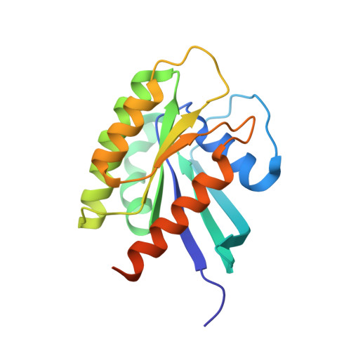
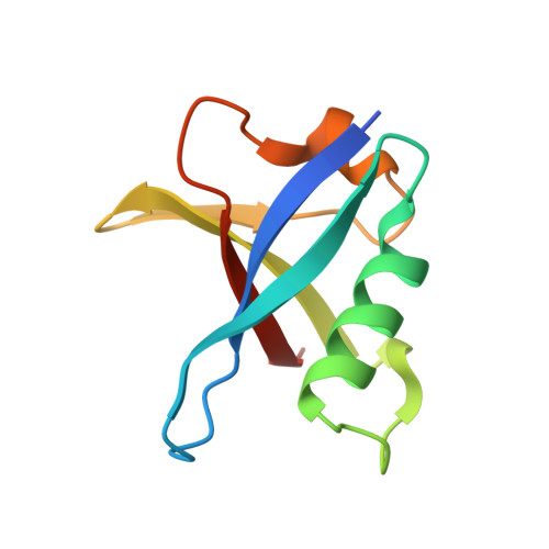
Structures of RGL1 RAS-Association Domain in Complex with KRAS and the Oncogenic G12V Mutant.

Eves, B.J.Gebregiworgis, T.Gasmi-Seabrook, G.M.C.Kuntz, D.A.Prive, G.G.Marshall, C.B.Ikura, M.

(2022) J Mol Biol 434: 167527-167527

  • DOI: https://doi.org/10.1016/j.jmb.2022.167527
  • Primary Citation of Related Structures:  
    7SCW, 7SCX

  • PubMed Abstract: 

    Ral Guanine Nucleotide Dissociation Stimulator Like 1 (RGL1) is a RAS effector protein that activates Ral GTPase by stimulating nucleotide exchange. Most structures of RAS-effector complexes are for the HRAS isoform; relatively few KRAS-effector structures have been solved, even though KRAS mutations are more frequent in human cancers. We determined crystal structures of KRAS/RGL1-RAS-association (RA) domain complexes and characterized the interaction in solution using nuclear magnetic resonance spectroscopy, size-exclusion chromatography combined with multi-angle light scattering and biolayer interferometry. We report structures of wild-type KRAS and the oncogenic G12V mutant in complex with the RA domain of RGL1 at < 2 Å resolution. KRAS WT /RGL1-RA crystallized as a 1:1 heterodimer, whilst KRAS G12V /RGL1-RA crystallized as a heterotetrameric structure in which RGL1-RA dimerized via domain-swapping the C-terminal beta-strand. Solution data indicated that KRAS WT and KRAS G12V in complex with RGL1-RA both exist predominantly as 1:1 dimers, while tetramerization occurs through very slow association. Through detailed structural analyses, the distance and angle between RAS α1 helix and RBD/RA α1 helix were found to differ significantly among RAS and RBD/RA complexes. The KRAS/RGL1-RA structures possess some of the largest α1 RAS /α1 Effector distances (21.7-22.2 Å), whereas the corresponding distances in previously reported RAS/RAF complexes are significantly shorter (15.2-17.7 Å). Contact map analysis identified unique structural signatures involving contacts between the β1-β2 loop of RA and the α1 helix of RAS, clearly distinguishing the KRAS/RGL1-RA (and other RAS/RA complexes) from RAS/RBD complexes. These results demonstrate that RAS effectors employ an assortment of finely-tuned docking surfaces to achieve optimal interactions with RAS.


  • Organizational Affiliation

    Princess Margaret Cancer Centre, University Health Network, Toronto, ON M5G 1L7, Canada. Electronic address: https://twitter.com/@B_Eves.


Macromolecules
Find similar proteins by:  (by identity cutoff)  |  3D Structure
Entity ID: 1
MoleculeChains Sequence LengthOrganismDetailsImage
Isoform 2B of GTPase KRas190Homo sapiensMutation(s): 1 
Gene Names: KRASKRAS2RASK2
EC: 3.6.5.2
UniProt & NIH Common Fund Data Resources
Find proteins for P01116 (Homo sapiens)
Explore P01116 
Go to UniProtKB:  P01116
PHAROS:  P01116
GTEx:  ENSG00000133703 
Entity Groups  
Sequence Clusters30% Identity50% Identity70% Identity90% Identity95% Identity100% Identity
UniProt GroupP01116
Sequence Annotations
Expand
  • Reference Sequence
Find similar proteins by:  (by identity cutoff)  |  3D Structure
Entity ID: 2
MoleculeChains Sequence LengthOrganismDetailsImage
Ral guanine nucleotide dissociation stimulator-like 193Homo sapiensMutation(s): 0 
Gene Names: RGL1KIAA0959RGL
UniProt & NIH Common Fund Data Resources
Find proteins for Q9NZL6 (Homo sapiens)
Explore Q9NZL6 
Go to UniProtKB:  Q9NZL6
PHAROS:  Q9NZL6
GTEx:  ENSG00000143344 
Entity Groups  
Sequence Clusters30% Identity50% Identity70% Identity90% Identity95% Identity100% Identity
UniProt GroupQ9NZL6
Sequence Annotations
Expand
  • Reference Sequence
Experimental Data & Validation

Experimental Data

  • Method: X-RAY DIFFRACTION
  • Resolution: 1.98 Å
  • R-Value Free:  0.192 (Depositor), 0.190 (DCC) 
  • R-Value Work:  0.156 (Depositor), 0.150 (DCC) 
  • R-Value Observed: 0.158 (Depositor) 
Space Group: P 63 2 2
Unit Cell:
Length ( Å )Angle ( ˚ )
a = 91.064α = 90
b = 91.064β = 90
c = 132.896γ = 120
Software Package:
Software NamePurpose
PHENIXrefinement
Aimlessdata scaling
XDSdata reduction
PHASERphasing

Structure Validation

View Full Validation Report


Ligand Structure Quality Assessment 

Created with Raphaël 2.3.0Worse 01 BetterLigand structure goodness of fit to experimental dataBest fitted GSPClick on this verticalbar to view details

Entry History & Funding Information

Deposition Data


Funding OrganizationLocationGrant Number
Not funded--

Revision History  (Full details and data files)

  • Version 1.0: 2022-05-18
    Type: Initial release
  • Version 1.1: 2022-11-30
    Changes: Database references
  • Version 1.2: 2023-10-25
    Changes: Data collection, Refinement description