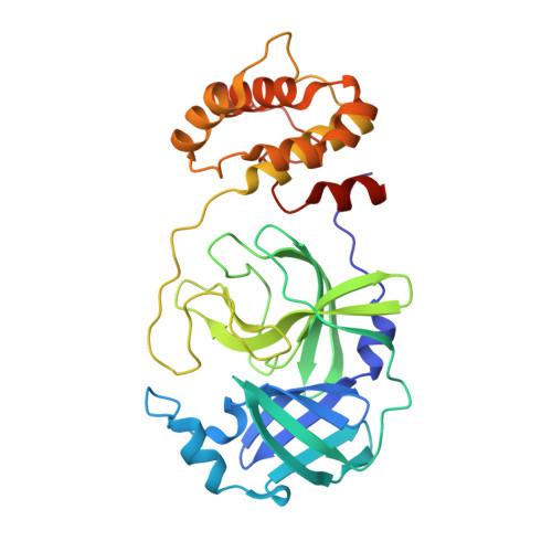
7XAX

Crystal structure of SARS coronavirus main protease in complex with Baicalei


Experimental Data Snapshot

  • Method: X-RAY DIFFRACTION
  • Resolution: 2.25 Å
  • R-Value Free: 0.280 
  • R-Value Work: 0.229 
  • R-Value Observed: 0.232 

Starting Model: experimental
View more details

wwPDB Validation   3D Report Full Report


Ligand Structure Quality Assessment 


This is version 1.2 of the entry. See complete history


Literature

Crystal structure of SARS-CoV 3C-like protease with baicalein.

Feng, J.Li, D.Zhang, J.Yin, X.Li, J.

(2022) Biochem Biophys Res Commun 611: 190-194

  • DOI: https://doi.org/10.1016/j.bbrc.2022.04.086
  • Primary Citation of Related Structures:  
    7XAX

  • PubMed Abstract: 

    The 3C-like protease (M pro , 3CL pro ) plays a key role in the replication process in coronaviruses (CoVs). The M pro is an essential enzyme mediates CoVs replication and is a promising target for development of antiviral drugs. Until now, baicalein has been shown the specific activity for SARS-CoV M pro in vitro experiments. In this study, we resolved the SARS-CoV M pro with baicalein by X-ray diffraction at 2.25 Å (PDB code 7XAX), which provided a structural basis for the research and development of baicalein as an anti-CoVs drug.


  • Organizational Affiliation

    Applied Biology Laboratory, College of Pharmaceutical and Biological Engineering, Shenyang University of Chemical Technology, Shenyang, 110142, China.


Macromolecules
Find similar proteins by:  (by identity cutoff)  |  3D Structure
Entity ID: 1
MoleculeChains Sequence LengthOrganismDetailsImage
3C-like proteinase nsp5
A, B
306Severe acute respiratory syndrome-related coronavirusMutation(s): 0 
EC: 3.4.22.69
UniProt
Find proteins for P0C6X7 (Severe acute respiratory syndrome coronavirus)
Explore P0C6X7 
Go to UniProtKB:  P0C6X7
Entity Groups  
Sequence Clusters30% Identity50% Identity70% Identity90% Identity95% Identity100% Identity
UniProt GroupP0C6X7
Sequence Annotations
Expand
  • Reference Sequence
Small Molecules
Ligands 1 Unique
IDChains Name / Formula / InChI Key2D Diagram3D Interactions
3WL (Subject of Investigation/LOI)
Query on 3WL

Download Ideal Coordinates CCD File 
C [auth A]5,6,7-trihydroxy-2-phenyl-4H-chromen-4-one
C15 H10 O5
FXNFHKRTJBSTCS-UHFFFAOYSA-N
Experimental Data & Validation

Experimental Data

  • Method: X-RAY DIFFRACTION
  • Resolution: 2.25 Å
  • R-Value Free: 0.280 
  • R-Value Work: 0.229 
  • R-Value Observed: 0.232 
  • Space Group: P 1
Unit Cell:
Length ( Å )Angle ( ˚ )
a = 55.51α = 90.951
b = 60.55β = 102.715
c = 68.28γ = 108.649
Software Package:
Software NamePurpose
PHENIXrefinement
XDSdata scaling
PHENIXphasing

Structure Validation

View Full Validation Report


Ligand Structure Quality Assessment 


Entry History & Funding Information

Deposition Data


Funding OrganizationLocationGrant Number
Not funded--

Revision History  (Full details and data files)

  • Version 1.0: 2023-03-22
    Type: Initial release
  • Version 1.1: 2023-11-29
    Changes: Data collection, Refinement description
  • Version 1.2: 2024-10-02
    Changes: Database references