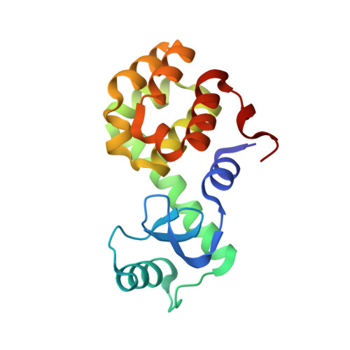
7XE6

T4 lysozyme mutant-S44C/C54T/N68C/A93C/C97A/T115C, pH7


Experimental Data Snapshot

  • Method: X-RAY DIFFRACTION
  • Resolution: 1.10 Å
  • R-Value Free: 0.168 
  • R-Value Work: 0.148 
  • R-Value Observed: 0.149 

Starting Model: experimental
View more details

wwPDB Validation   3D Report Full Report


This is version 1.2 of the entry. See complete history


Literature

Creation of Cross-Linked Crystals With Intermolecular Disulfide Bonds Connecting Symmetry-Related Molecules Allows Retention of Tertiary Structure in Different Solvent Conditions.

Hiromoto, T.Ikura, T.Honjo, E.Blaber, M.Kuroki, R.Tamada, T.

(2022) Front Mol Biosci 9: 908394-908394

  • DOI: https://doi.org/10.3389/fmolb.2022.908394
  • Primary Citation of Related Structures:  
    7XE5, 7XE6, 7XE7, 7XE9, 7XEA

  • PubMed Abstract: 

    Protein crystals are generally fragile and sensitive to subtle changes such as pH, ionic strength, and/or temperature in their crystallization mother liquor. Here, using T4 phage lysozyme as a model protein, the three-dimensional rigidification of protein crystals was conducted by introducing disulfide cross-links between neighboring molecules in the crystal. The effect of cross-linking on the stability of the crystals was evaluated by microscopic observation and X-ray diffraction. When soaking the obtained cross-linked crystals into a precipitant-free solution, the crystals held their shape without dissolution and diffracted to approximately 1.1 Å resolution, comparable to that of the non-cross-linked crystals. Such cross-linked crystals maintained their diffraction even when immersed in other solutions with pH values from 4 to 10, indicating that the disulfide cross-linking made the packing contacts enforced and resulted in some mechanical strength in response to changes in the preservation conditions. Furthermore, the cross-linked crystals gained stability to permit soaking into solutions containing high concentrations of organic solvents. The results suggest the possibility of obtaining protein crystals for effective drug screening by introducing appropriate cross-linked disulfide bonds.


  • Organizational Affiliation

    Institute for Quantum Life Science, National Institutes for Quantum Science and Technology, Ibaraki, Japan.


Macromolecules
Find similar proteins by:  (by identity cutoff)  |  3D Structure
Entity ID: 1
MoleculeChains Sequence LengthOrganismDetailsImage
Endolysin164Tequatrovirus T4Mutation(s): 6 
Gene Names: eT4Tp126
EC: 3.2.1.17
UniProt
Find proteins for D9IEF7 (Enterobacteria phage T4)
Explore D9IEF7 
Go to UniProtKB:  D9IEF7
Entity Groups  
Sequence Clusters30% Identity50% Identity70% Identity90% Identity95% Identity100% Identity
UniProt GroupD9IEF7
Sequence Annotations
Expand
  • Reference Sequence
Small Molecules
Ligands 3 Unique
IDChains Name / Formula / InChI Key2D Diagram3D Interactions
HEZ
Query on HEZ

Download Ideal Coordinates CCD File 
B [auth A],
C [auth A]
HEXANE-1,6-DIOL
C6 H14 O2
XXMIOPMDWAUFGU-UHFFFAOYSA-N
GOL
Query on GOL

Download Ideal Coordinates CCD File 
D [auth A],
E [auth A]
GLYCEROL
C3 H8 O3
PEDCQBHIVMGVHV-UHFFFAOYSA-N
NA
Query on NA

Download Ideal Coordinates CCD File 
F [auth A],
G [auth A],
H [auth A]
SODIUM ION
Na
FKNQFGJONOIPTF-UHFFFAOYSA-N
Experimental Data & Validation

Experimental Data

  • Method: X-RAY DIFFRACTION
  • Resolution: 1.10 Å
  • R-Value Free: 0.168 
  • R-Value Work: 0.148 
  • R-Value Observed: 0.149 
  • Space Group: P 32 2 1
Unit Cell:
Length ( Å )Angle ( ˚ )
a = 59.999α = 90
b = 59.999β = 90
c = 97.101γ = 120
Software Package:
Software NamePurpose
PHENIXrefinement
HKL-2000data reduction
HKL-2000data scaling

Structure Validation

View Full Validation Report


Entry History & Funding Information

Deposition Data


Funding OrganizationLocationGrant Number
Not funded--

Revision History  (Full details and data files)

  • Version 1.0: 2023-03-22
    Type: Initial release
  • Version 1.1: 2023-11-29
    Changes: Data collection, Refinement description
  • Version 1.2: 2024-11-13
    Changes: Structure summary