


The surface-exposed lipo-protein of BtuG2 in complex with cyanocobalamin.
Whittaker, J., Guskov, A.To be published.
Experimental Data Snapshot
Starting Model: experimental
View more details
Entity ID: 1 | |||||
|---|---|---|---|---|---|
| Molecule | Chains | Sequence Length | Organism | Details | Image |
| Surface layer protein | 321 | Bacteroides thetaiotaomicron | Mutation(s): 0 Gene Names: BT_1954 | 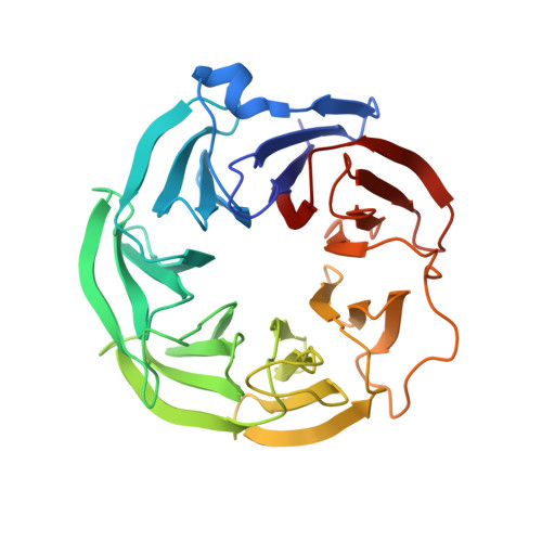 | |
UniProt | |||||
Entity Groups | |||||
| Sequence Clusters | 30% Identity50% Identity70% Identity90% Identity95% Identity100% Identity | ||||
| UniProt Group | Q8A6D0 | ||||
Sequence AnnotationsExpand | |||||
Reference Sequence | |||||
| Ligands 5 Unique | |||||
|---|---|---|---|---|---|
| ID | Chains | Name / Formula / InChI Key | 2D Diagram | 3D Interactions | |
| CNC Download:Ideal Coordinates CCD File | AA [auth C], GA [auth D], L [auth A], T [auth B] | CYANOCOBALAMIN C63 H89 Co N14 O14 P SYZBZQWSWIJYAR-UVKKECPRSA-M |  | ||
| PEG Download:Ideal Coordinates CCD File | U [auth B], Y [auth C] | DI(HYDROXYETHYL)ETHER C4 H10 O3 MTHSVFCYNBDYFN-UHFFFAOYSA-N |  | ||
| SO4 Download:Ideal Coordinates CCD File | S [auth B], Z [auth C] | SULFATE ION O4 S QAOWNCQODCNURD-UHFFFAOYSA-L |  | ||
| GOL Download:Ideal Coordinates CCD File | BA [auth D] CA [auth D] DA [auth D] E [auth A] EA [auth D] | GLYCEROL C3 H8 O3 PEDCQBHIVMGVHV-UHFFFAOYSA-N |  | ||
| NA Download:Ideal Coordinates CCD File | HA [auth D], IA [auth D] | SODIUM ION Na FKNQFGJONOIPTF-UHFFFAOYSA-N |  | ||
| Length ( Å ) | Angle ( ˚ ) |
|---|---|
| a = 49.24 | α = 89.97 |
| b = 79.936 | β = 89.89 |
| c = 101.047 | γ = 82.28 |
| Software Name | Purpose |
|---|---|
| REFMAC | refinement |
| Aimless | data scaling |
| PDB_EXTRACT | data extraction |
| XDS | data reduction |
| PHASER | phasing |
| Funding Organization | Location | Grant Number |
|---|---|---|
| Not funded | Netherlands | -- |


