8H9D | pdb_00008h9d

Crystal structure of Cas12a protein


Experimental Data Snapshot

  • Method: X-RAY DIFFRACTION
  • Resolution: 3.10 Å
  • R-Value Free: 
    0.252 (Depositor), 0.280 (DCC) 
  • R-Value Work: 
    0.232 (Depositor) 
  • R-Value Observed: 
    0.233 (Depositor) 

Starting Model: experimental
View more details

wwPDB Validation 3D Report Full Report

Validation slider image for 8H9D

This is version 1.2 of the entry. See complete history

Literature

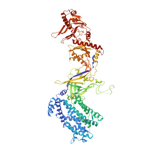
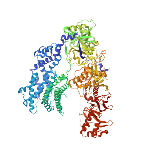
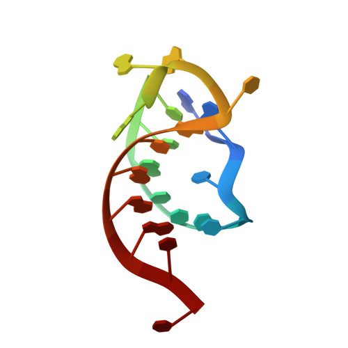
Structures of apo Cas12a and its complex with crRNA and DNA reveal the dynamics of ternary complex formation and target DNA cleavage.

Jianwei, L.Jobichen, C.Machida, S.Meng, S.Read, R.J.Hongying, C.Jian, S.Yuan, Y.A.Sivaraman, J.

(2023) PLoS Biol 21: e3002023-e3002023

  • DOI: https://doi.org/10.1371/journal.pbio.3002023
  • Primary Citation Related Structures: 
    8H9D, 8I54

  • PubMed Abstract: 

    Cas12a is a programmable nuclease for adaptive immunity against invading nucleic acids in CRISPR-Cas systems. Here, we report the crystal structures of apo Cas12a from Lachnospiraceae bacterium MA2020 (Lb2) and the Lb2Cas12a+crRNA complex, as well as the cryo-EM structure and functional studies of the Lb2Cas12a+crRNA+DNA complex. We demonstrate that apo Lb2Cas12a assumes a unique, elongated conformation, whereas the Lb2Cas12a+crRNA binary complex exhibits a compact conformation that subsequently rearranges to a semi-open conformation in the Lb2Cas12a+crRNA+DNA ternary complex. Notably, in solution, apo Lb2Cas12a is dynamic and can exist in both elongated and compact forms. Residues from Met493 to Leu523 of the WED domain undergo major conformational changes to facilitate the required structural rearrangements. The REC lobe of Lb2Cas12a rotates 103° concomitant with rearrangement of the hinge region close to the WED and RuvC II domains to position the RNA-DNA duplex near the catalytic site. Our findings provide insight into crRNA recognition and the mechanism of target DNA cleavage.


  • Organizational Affiliation
    • Department of Biological Sciences, National University of Singapore, Singapore, Singapore.

Macromolecule Content 

  • Total Structure Weight: 289.32 kDa 
  • Atom Count: 16,986 
  • Modeled Residue Count: 2,081 
  • Deposited Residue Count: 2,434 
  • Unique protein chains: 2
  • Unique nucleic acid chains: 1

Macromolecules


Find similar proteins by:|  3D Structure
Entity ID: 1
MoleculeChains  Sequence LengthOrganismDetailsImage
Cas12AA [auth B]1,206Lachnospiraceae bacteriumMutation(s): 0 
Find similar proteins by:|  3D Structure
Entity ID: 2
MoleculeChains  Sequence LengthOrganismDetailsImage
Cas12AB [auth A]1,207Lachnospiraceae bacteriumMutation(s): 0 
Find similar nucleic acids by:  Sequence
Entity ID: 3
MoleculeChains LengthOrganismImage
RNA (5'-R(P*AP*AP*UP*UP*UP*CP*UP*AP*CP*UP*AP*AP*GP*UP*GP*UP*AP*GP*AP*UP*C)-3')C [auth G]21Lachnospiraceae bacterium
Sequence Annotations
Expand
Reference Sequence

Experimental Data & Validation

Experimental Data

  • Method: X-RAY DIFFRACTION
  • Resolution: 3.10 Å
  • R-Value Free:  0.252 (Depositor), 0.280 (DCC) 
  • R-Value Work:  0.232 (Depositor) 
  • R-Value Observed: 0.233 (Depositor) 
Space Group: C 1 2 1
Unit Cell:
Length ( Å )Angle ( ˚ )
a = 235.701α = 90
b = 85.628β = 110.15
c = 139.303γ = 90
Software Package:
Software NamePurpose
PHENIXrefinement
HKL-2000data reduction
HKL-2000data scaling
PHASERphasing

Structure Validation

View Full Validation Report


Entry History 

& Funding Information

Deposition Data


Funding OrganizationLocationGrant Number
Ministry of Education (MoE, Singapore)SingaporeR-154-000-C07-114
Ministry of Education (MoE, Singapore)SingaporeR154-000-A39-112

Revision History  (Full details and data files)

  • Version 1.0: 2023-02-08
    Type: Initial release
  • Version 1.1: 2023-08-16
    Changes: Data collection, Database references
  • Version 1.2: 2023-11-29
    Changes: Refinement description