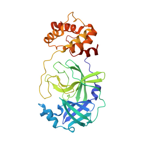
8HQF

Crystal structure of SARS-Cov-2 main protease G15S mutant in complex with inhibitor YH-53


Experimental Data Snapshot

  • Method: X-RAY DIFFRACTION
  • Resolution: 1.51 Å
  • R-Value Free: 0.230 
  • R-Value Work: 0.208 
  • R-Value Observed: 0.209 

Starting Model: experimental
View more details

wwPDB Validation   3D Report Full Report


Ligand Structure Quality Assessment 


This is version 1.1 of the entry. See complete history


Literature

Crystal structures of main proteases of SARS-CoV-2 variants bound to a benzothiazole-based inhibitor.

Luo, J.Wang, W.Jiang, H.Li, W.Zeng, P.Wang, J.Zhou, X.Zou, X.Chen, S.Wang, Q.Zhang, J.Li, J.

(2023) Acta Biochim Biophys Sin (Shanghai) 55: 1257-1264

  • DOI: https://doi.org/10.3724/abbs.2023053
  • Primary Citation of Related Structures:  
    8HQF, 8HQG, 8HQH, 8HQI, 8HQJ

  • PubMed Abstract: 

    Main protease (M pro ) serves as an indispensable factor in the life cycle of severe acute respiratory syndrome coronavirus 2 (SARS-CoV-2) as well as its constantly emerging variants and is therefore considered an attractive target for antiviral drug development. Benzothiazole-based inhibitors targeting M pro have recently been investigated by several groups and proven to be promising leads for coronaviral drug development. In the present study, we determine the crystal structures of a benzothiazole-based inhibitor, YH-53, bound to M pro mutants from SARS-CoV-2 variants of concern (VOCs) or variants of interest (VOIs), including K90R (Beta, B.1.351), G15S (Lambda, C.37), Y54C (Delta, AY.4), M49I (Omicron, BA.5) and P132H (Omicron, B.1.1.529). The structures show that the benzothiazole group in YH-53 forms a C-S covalent bond with the sulfur atom of catalytic residue Cys145 in SARS-CoV-2 M pro mutants. Structural analysis reveals the key molecular determinants necessary for interaction and illustrates the binding mode of YH-53 to these mutant M pro s. In conclusion, structural insights from this study offer more information to develop benzothiazole-based drugs that are broader spectrum, more effective and safer.


  • Organizational Affiliation

    College of Pharmaceutical Sciences, Gannan Medical University, Ganzhou 341000, China.


Macromolecules
Find similar proteins by:  (by identity cutoff)  |  3D Structure
Entity ID: 1
MoleculeChains Sequence LengthOrganismDetailsImage
Non-structural protein 7
A, B
299Severe acute respiratory syndrome coronavirus 2Mutation(s): 1 
Gene Names: rep1a-1b
UniProt
Find proteins for P0DTD1 (Severe acute respiratory syndrome coronavirus 2)
Explore P0DTD1 
Go to UniProtKB:  P0DTD1
Entity Groups  
Sequence Clusters30% Identity50% Identity70% Identity90% Identity95% Identity100% Identity
UniProt GroupP0DTD1
Sequence Annotations
Expand
  • Reference Sequence
Small Molecules
Ligands 1 Unique
IDChains Name / Formula / InChI Key2D Diagram3D Interactions
HUR (Subject of Investigation/LOI)
Query on HUR

Download Ideal Coordinates CCD File 
C [auth A],
D [auth B]
N-[(2S)-1-[[(2S)-1-(1,3-benzothiazol-2-yl)-1-oxidanylidene-3-[(3S)-2-oxidanylidenepyrrolidin-3-yl]propan-2-yl]amino]-4-methyl-1-oxidanylidene-pentan-2-yl]-4-methoxy-1H-indole-2-carboxamide
C30 H33 N5 O5 S
JBLLRCOZJMVOAE-HSQYWUDLSA-N
Experimental Data & Validation

Experimental Data

  • Method: X-RAY DIFFRACTION
  • Resolution: 1.51 Å
  • R-Value Free: 0.230 
  • R-Value Work: 0.208 
  • R-Value Observed: 0.209 
  • Space Group: P 1 21 1
Unit Cell:
Length ( Å )Angle ( ˚ )
a = 55.381α = 90
b = 99.44β = 108.253
c = 59.461γ = 90
Software Package:
Software NamePurpose
PHENIXrefinement
XDSdata reduction
XDSdata scaling
PHENIXphasing

Structure Validation

View Full Validation Report


Ligand Structure Quality Assessment 


Entry History & Funding Information

Deposition Data


Funding OrganizationLocationGrant Number
Not funded--

Revision History  (Full details and data files)

  • Version 1.0: 2024-01-31
    Type: Initial release
  • Version 1.1: 2025-01-01
    Changes: Database references, Structure summary