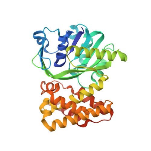
Implication of Molecular Constraints Facilitating the Functional Evolution of Pseudomonas aeruginosa KPR2 into a Versatile alpha-Keto-Acid Reductase.
Basu Choudhury, G., Datta, S.(2024) Biochemistry 63: 1808-1823
- PubMed: 38962820
- DOI: https://doi.org/10.1021/acs.biochem.4c00087
- Primary Citation of Related Structures:
8IWG, 8IWQ, 8IX9, 8IXH, 8IXM - PubMed Abstract:
Theoretical concepts linking the structure, function, and evolution of a protein, while often intuitive, necessitate validation through investigations in real-world systems. Our study empirically explores the evolutionary implications of multiple gene copies in an organism by shedding light on the structure-function modulations observed in Pseudomonas aeruginosa 's second copy of ketopantoate reductase (PaKPR2). We demonstrated with two apo structures that the typical active site cleft of the protein transforms into a two-sided pocket where a molecular gate made up of two residues controls the substrate entry site, resulting in its inactivity toward the natural substrate ketopantoate. Strikingly, this structural modification made the protein active against several important α-keto-acid substrates with varied efficiency. Structural constraints at the binding site for this altered functional trait were analyzed with two binary complexes that show the conserved residue microenvironment faces restricted movements due to domain closure. Finally, its mechanistic highlights gathered from a ternary complex structure help in delineating the molecular perspectives behind its kinetic cooperativity toward these broad range of substrates. Detailed structural characteristics of the protein presented here also identified four key amino acid residues responsible for its versatile α-keto-acid reductase activity, which can be further modified to improve its functional properties through protein engineering.
Organizational Affiliation:
CSIR-Indian Institute of Chemical Biology, Raja S C Mullick Road, Jadavpur, Kolkata 700032, India.


