8J4F | pdb_00008j4f

Structure of human Nav1.7 in complex with Hardwickii acid


Experimental Data Snapshot

  • Method: ELECTRON MICROSCOPY
  • Resolution: 3.00 Å
  • Aggregation State: PARTICLE 
  • Reconstruction Method: SINGLE PARTICLE 

wwPDB Validation 3D Report Full Report

Validation slider image for 8J4F

This is version 1.1 of the entry. See complete history

Literature

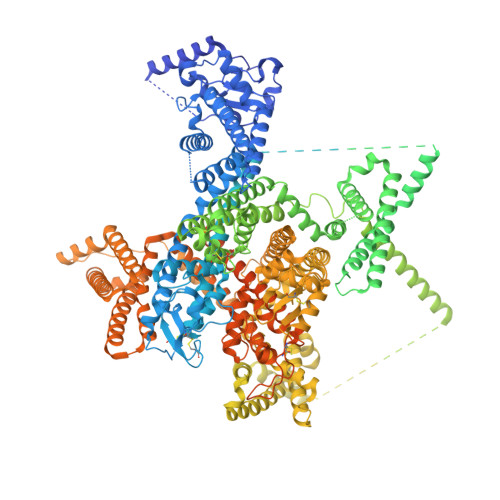
Structural mapping of Na v 1.7 antagonists.

Wu, Q.Huang, J.Fan, X.Wang, K.Jin, X.Huang, G.Li, J.Pan, X.Yan, N.

(2023) Nat Commun 14: 3224-3224

  • DOI: https://doi.org/10.1038/s41467-023-38942-3
  • Primary Citation Related Structures: 
    8I5B, 8I5G, 8I5X, 8I5Y, 8J4F, 8S9B, 8S9C

  • PubMed Abstract: 

    Voltage-gated sodium (Na v ) channels are targeted by a number of widely used and investigational drugs for the treatment of epilepsy, arrhythmia, pain, and other disorders. Despite recent advances in structural elucidation of Na v channels, the binding mode of most Na v -targeting drugs remains unknown. Here we report high-resolution cryo-EM structures of human Na v 1.7 treated with drugs and lead compounds with representative chemical backbones at resolutions of 2.6-3.2 Å. A binding site beneath the intracellular gate (site BIG) accommodates carbamazepine, bupivacaine, and lacosamide. Unexpectedly, a second molecule of lacosamide plugs into the selectivity filter from the central cavity. Fenestrations are popular sites for various state-dependent drugs. We show that vinpocetine, a synthetic derivative of a vinca alkaloid, and hardwickiic acid, a natural product with antinociceptive effect, bind to the III-IV fenestration, while vixotrigine, an analgesic candidate, penetrates the IV-I fenestration of the pore domain. Our results permit building a 3D structural map for known drug-binding sites on Na v channels summarized from the present and previous structures.


  • Organizational Affiliation
    • Beijing Frontier Research Center for Biological Structures, Tsinghua-Peking Joint Center for Life Sciences, School of Life Sciences, Tsinghua University, Beijing, 100084, China.

Macromolecule Content 

  • Total Structure Weight: 268.61 kDa 
  • Atom Count: 12,419 
  • Modeled Residue Count: 1,446 
  • Deposited Residue Count: 2,246 
  • Unique protein chains: 2

Macromolecules

Find similar proteins by:|  3D Structure
Entity ID: 1
MoleculeChains  Sequence LengthOrganismDetailsImage
Sodium channel protein type 9 subunit alpha2,028Homo sapiensMutation(s): 0 
Gene Names: SCN9ANENA
Membrane Entity: Yes 
UniProt & NIH Common Fund Data Resources
Find proteins for Q15858 (Homo sapiens)
Explore Q15858 
Go to UniProtKB:  Q15858
PHAROS:  Q15858
GTEx:  ENSG00000169432 
Entity Groups
Sequence Clusters30% Identity50% Identity70% Identity90% Identity95% Identity100% Identity
UniProt GroupQ15858
Glycosylation
Glycosylation Sites: 4Go to GlyGen: Q15858-1
Sequence Annotations
Expand
Reference Sequence
Find similar proteins by:|  3D Structure
Entity ID: 2
MoleculeChains  Sequence LengthOrganismDetailsImage
Sodium channel subunit beta-1218Homo sapiensMutation(s): 0 
Gene Names: SCN1B
Membrane Entity: Yes 
UniProt & NIH Common Fund Data Resources
Find proteins for Q07699 (Homo sapiens)
Explore Q07699 
Go to UniProtKB:  Q07699
PHAROS:  Q07699
GTEx:  ENSG00000105711 
Entity Groups
Sequence Clusters30% Identity50% Identity70% Identity90% Identity95% Identity100% Identity
UniProt GroupQ07699
Glycosylation
Glycosylation Sites: 4Go to GlyGen: Q07699-1
Sequence Annotations
Expand
Reference Sequence

Oligosaccharides

Help  
Entity ID: 3
MoleculeChains Length2D Diagram GlycosylationD Interactions
2-acetamido-2-deoxy-beta-D-glucopyranose-(1-4)-2-acetamido-2-deoxy-beta-D-glucopyranose
C, D, E
2N-Glycosylation
Glycosylation Resources
GlyTouCan: G42666HT
GlyCosmos: G42666HT
GlyGen: G42666HT

Small Molecules

Ligands 6 Unique
IDChains Name / Formula / InChI Key2D Diagram3D Interactions
PCW

Query on PCW


Download:Ideal Coordinates CCD File
M [auth A],
P [auth A],
Q [auth A],
X [auth A]
1,2-DIOLEOYL-SN-GLYCERO-3-PHOSPHOCHOLINE
C44 H85 N O8 P
SNKAWJBJQDLSFF-NVKMUCNASA-O
9Z9

Query on 9Z9


Download:Ideal Coordinates CCD File
K [auth A](3beta,14beta,17beta,25R)-3-[4-methoxy-3-(methoxymethyl)butoxy]spirost-5-en
C34 H56 O5
CEEBZAXXSRFQIC-GZSGZGDASA-N
LPE

Query on LPE


Download:Ideal Coordinates CCD File
CA [auth B]
L [auth A]
N [auth A]
O [auth A]
R [auth A]
CA [auth B],
L [auth A],
N [auth A],
O [auth A],
R [auth A],
S [auth A],
T [auth A],
U [auth A],
V [auth A],
W [auth A]
1-O-OCTADECYL-SN-GLYCERO-3-PHOSPHOCHOLINE
C26 H57 N O6 P
XKBJVQHMEXMFDZ-AREMUKBSSA-O
Y01

Query on Y01


Download:Ideal Coordinates CCD File
I [auth A],
J [auth A],
Y [auth A]
CHOLESTEROL HEMISUCCINATE
C31 H50 O4
WLNARFZDISHUGS-MIXBDBMTSA-N
UK0

Query on UK0


Download:Ideal Coordinates CCD File
H [auth A](4~{a}~{R},5~{S},6~{R},8~{a}~{R})-5-[2-(furan-3-yl)ethyl]-5,6,8~{a}-trimethyl-3,4,4~{a},6,7,8-hexahydronaphthalene-1-carboxylic acid
C20 H28 O3
HHWOKJDCJVESIF-JBCDFXQESA-N
NAG

Query on NAG


Download:Ideal Coordinates CCD File
AA [auth B],
BA [auth B],
F [auth A],
G [auth A],
Z [auth B]
2-acetamido-2-deoxy-beta-D-glucopyranose
C8 H15 N O6
OVRNDRQMDRJTHS-FMDGEEDCSA-N

Experimental Data & Validation

Experimental Data

  • Method: ELECTRON MICROSCOPY
  • Resolution: 3.00 Å
  • Aggregation State: PARTICLE 
  • Reconstruction Method: SINGLE PARTICLE 
EM Software:
TaskSoftware PackageVersion
MODEL REFINEMENTPHENIX1.20_4459:

Structure Validation

View Full Validation Report


Entry History 

& Funding Information

Deposition Data


Funding OrganizationLocationGrant Number
National Natural Science Foundation of China (NSFC)China32271252

Revision History  (Full details and data files)

  • Version 1.0: 2023-06-14
    Type: Initial release
  • Version 1.1: 2024-10-30
    Changes: Data collection, Structure summary