Identification of fragments targeting SMYD3 using highly sensitive kinetic and multiplexed biosensor-based screening.
FitzGerald, E.A., Cederfelt, D., Lund, B.A., Myers, N.E.M., Zhang, H., Dobritzsch, D., Danielson, U.H.(2024) RSC Med Chem 15: 1982-1990
- PubMed: 38911161 Search on PubMedSearch on PubMed Central
- DOI: https://doi.org/10.1039/d4md00093e
- Primary Citation Related Structures:
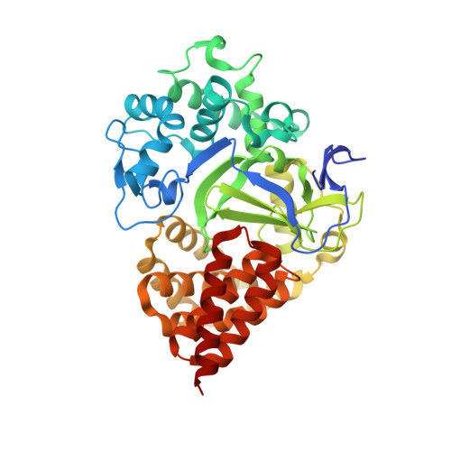
8OWO - PubMed Abstract:
A 1056-membered fragment library has been screened against SMYD3 using a novel multiplexed experimental design implemented in a grating coupled interferometry (GCI)-based biosensor. SMYD3 is a prospective target for anticancer drugs and the focus has initially been on discovery of inhibitors of its lysine methyl transferase activity. However, it has multiple protein interaction partners and several potential roles in carcinogenesis. It therefore remains unclear what mode of action ligands targeting the protein should have. Our goal was therefore to identify new ligands and discriminate hits that interact with the active site and those that interact with other sites. In addition, we were interested in selecting hits based on kinetic features rather than affinity. Screening was done in parallel against SMYD3 alone or SMYD3 with the active site blocked by a tight binding inhibitor. Hit selection was primarily based on dissociation rates. In total, 20 fragments were selected as hits, of which half apparently targeted the active site and half targeted other sites. Twelve of the hits were selected for structural analysis using X-ray crystallography in order to identify binding sites and modes of binding. Four of the hits were successfully identified in crystal structures with SMYD3; the others did not show any electron densities for ligands in the crystals. Although it might be possible to optimize the crystallography approach for a better success rate, it was clear that the sensitivity and time resolution of the biosensor assay was exceptional and enabled kinetic rate constants to be estimated for fragments. Fragments are typically considered to interact too rapidly for such quantification to be possible. This approach consequently represents a paradigm shift. In addition, the multiplexed approach allows ligands targeting different sites to be rationally selected already in the fragment library screening stage.
- Department of Chemistry - BMC, Uppsala University Uppsala Sweden helena.danielson@kemi.uu.se.
Organizational Affiliation: