9B59

Ubiquitin E2-Ub-E3 HECT tetrahedral transthiolation intermediate mimic - state 5


Experimental Data Snapshot

  • Method: ELECTRON MICROSCOPY
  • Resolution: 3.49 Å
  • Aggregation State: PARTICLE 
  • Reconstruction Method: SINGLE PARTICLE 

wwPDB Validation   3D Report Full Report


This is version 1.5 of the entry. See complete history


Literature

Structural basis for transthiolation intermediates in the ubiquitin pathway.

Kochanczyk, T.Hann, Z.S.Lux, M.C.Delos Reyes, A.M.V.Ji, C.Tan, D.S.Lima, C.D.

(2024) Nature 633: 216-223

  • DOI: https://doi.org/10.1038/s41586-024-07828-9
  • Primary Citation of Related Structures:  
    9B55, 9B56, 9B57, 9B58, 9B59, 9B5A, 9B5B, 9B5C, 9B5D, 9B5E, 9B5F, 9B5G, 9B5H, 9B5I, 9B5J, 9B5K, 9B5L, 9B5M, 9B5N, 9B5O, 9B5P, 9B5Q, 9B5R, 9B5S, 9B5T, 9B5U, 9B5V, 9B5W, 9B5X

  • PubMed Abstract: 

    Transthiolation (also known as transthioesterification) reactions are used in the biosynthesis of acetyl coenzyme A, fatty acids and polyketides, and for post-translational modification by ubiquitin (Ub) and ubiquitin-like (Ubl) proteins 1-3 . For the Ub pathway, E1 enzymes catalyse transthiolation from an E1~Ub thioester to an E2~Ub thioester. Transthiolation is also required for transfer of Ub from an E2~Ub thioester to HECT (homologous to E6AP C terminus) and RBR (ring-between-ring) E3 ligases to form E3~Ub thioesters 4-6 . How isoenergetic transfer of thioester bonds is driven forward by enzymes in the Ub pathway remains unclear. Here we isolate mimics of transient transthiolation intermediates for E1-Ub(T)-E2 and E2-Ub(T)-E3 HECT complexes (where T denotes Ub in a thioester or Ub undergoing transthiolation) using a chemical strategy with native enzymes and near-native Ub to capture and visualize a continuum of structures determined by single-particle cryo-electron microscopy. These structures and accompanying biochemical experiments illuminate conformational changes in Ub, E1, E2 and E3 that are coordinated with the chemical reactions to facilitate directional transfer of Ub from each enzyme to the next.


  • Organizational Affiliation

    Structural Biology Program, Sloan Kettering Institute, New York, NY, USA.


Macromolecules
Find similar proteins by:  (by identity cutoff)  |  3D Structure
Entity ID: 1
MoleculeChains Sequence LengthOrganismDetailsImage
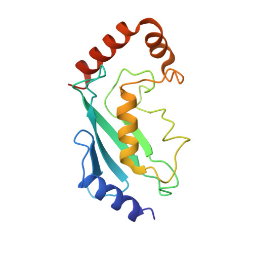
Ubiquitin-conjugating enzyme E2 4153Schizosaccharomyces pombe 972h-Mutation(s): 2 
Gene Names: ubc4SPBC119.02
EC: 2.3.2.23
UniProt
Find proteins for P46595 (Schizosaccharomyces pombe (strain 972 / ATCC 24843))
Explore P46595 
Go to UniProtKB:  P46595
Entity Groups  
Sequence Clusters30% Identity50% Identity70% Identity90% Identity95% Identity100% Identity
UniProt GroupP46595
Sequence Annotations
Expand
  • Reference Sequence
Find similar proteins by:  (by identity cutoff)  |  3D Structure
Entity ID: 2
MoleculeChains Sequence LengthOrganismDetailsImage
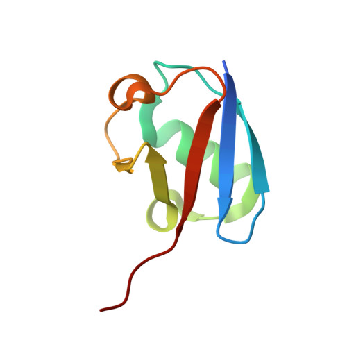
Ubiquitin79Schizosaccharomyces pombe 972h-Mutation(s): 0 
Gene Names: uep1ubi2SPAC1805.12c
UniProt
Find proteins for P0CH07 (Schizosaccharomyces pombe (strain 972 / ATCC 24843))
Explore P0CH07 
Go to UniProtKB:  P0CH07
Entity Groups  
Sequence Clusters30% Identity50% Identity70% Identity90% Identity95% Identity100% Identity
UniProt GroupP0CH07
Sequence Annotations
Expand
  • Reference Sequence
Find similar proteins by:  (by identity cutoff)  |  3D Structure
Entity ID: 3
MoleculeChains Sequence LengthOrganismDetailsImage
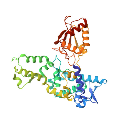
E3 ubiquitin-protein ligase pub2380Schizosaccharomyces pombe 972h-Mutation(s): 0 
Gene Names: pub2SPAC1805.15c
EC: 2.3.2.26
UniProt
Find proteins for Q9UTG2 (Schizosaccharomyces pombe (strain 972 / ATCC 24843))
Explore Q9UTG2 
Go to UniProtKB:  Q9UTG2
Entity Groups  
Sequence Clusters30% Identity50% Identity70% Identity90% Identity95% Identity100% Identity
UniProt GroupQ9UTG2
Sequence Annotations
Expand
  • Reference Sequence
Small Molecules
Ligands 1 Unique
IDChains Name / Formula / InChI Key2D Diagram3D Interactions
A1AIV (Subject of Investigation/LOI)
Query on A1AIV

Download Ideal Coordinates CCD File 
D [auth B]4-aminobutanenitrile
C4 H8 N2
XGYKKVTZDQDYJQ-UHFFFAOYSA-N
Experimental Data & Validation

Experimental Data

  • Method: ELECTRON MICROSCOPY
  • Resolution: 3.49 Å
  • Aggregation State: PARTICLE 
  • Reconstruction Method: SINGLE PARTICLE 
EM Software:
TaskSoftware PackageVersion
MODEL REFINEMENTPHENIX1.20.1_4487:
RECONSTRUCTIONcryoSPARC

Structure Validation

View Full Validation Report


Entry History & Funding Information

Deposition Data


Funding OrganizationLocationGrant Number
Howard Hughes Medical Institute (HHMI)United States--
National Institutes of Health/National Institute of General Medical Sciences (NIH/NIGMS)United StatesR35GM118080

Revision History  (Full details and data files)

  • Version 1.0: 2024-06-05
    Type: Initial release
  • Version 1.1: 2024-08-14
    Changes: Data collection, Database references
  • Version 1.2: 2024-08-28
    Changes: Data collection, Database references
  • Version 1.3: 2024-09-04
    Changes: Data collection, Database references
  • Version 1.4: 2024-09-11
    Changes: Data collection, Database references
  • Version 1.5: 2024-09-18
    Changes: Data collection