9BOX

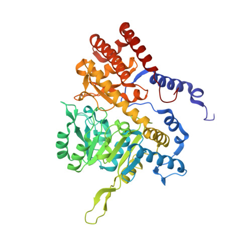
Room-temperature X-ray structure of human mitochondrial serine hydroxymethyltransferase (hSHMT2) with PLP-glycine external aldimine and 5-formyltetrahydrofolate (folinic acid)


Experimental Data Snapshot

  • Method: X-RAY DIFFRACTION
  • Resolution: 2.10 Å
  • R-Value Free: 0.189 
  • R-Value Work: 0.162 
  • R-Value Observed: 0.164 

Starting Model: experimental
View more details

wwPDB Validation   3D Report Full Report


Ligand Structure Quality Assessment 


This is version 1.0 of the entry. See complete history


Literature

Universality of critical active site glutamate as an acid-base catalyst in serine hydroxymethyltransferase function.

Drago, V.N.Phillips, R.S.Kovalevsky, A.

(2024) Chem Sci 15: 12827-12844

  • DOI: https://doi.org/10.1039/d4sc03187c
  • Primary Citation of Related Structures:  
    9BOH, 9BOW, 9BOX, 9BPE

  • PubMed Abstract: 

    Serine hydroxymethyltransferase (SHMT) is a key enzyme in the one-carbon metabolic pathway, utilizing the vitamin B 6 derivative pyridoxal 5'-phosphate (PLP) and vitamin B 9 derivative tetrahydrofolate (THF) coenzymes to produce essential biomolecules. Many types of cancer utilize SHMT in metabolic reprogramming, exposing the enzyme as a compelling target for antimetabolite chemotherapies. In pursuit of elucidating the catalytic mechanism of SHMT to aid in the design of SHMT-specific inhibitors, we have used room-temperature neutron crystallography to directly determine the protonation states in a model enzyme Thermus thermophilus SHMT ( Tth SHMT), which exhibits a conserved active site compared to human mitochondrial SHMT2 (hSHMT2). Here we report the analysis of Tth SHMT, with PLP in the internal aldimine form and bound THF-analog, folinic acid (FA), by neutron crystallography to reveal H atom positions in the active site, including PLP and FA. We observed protonated catalytic Glu53 revealing its ability to change protonation state upon FA binding. Furthermore, we obtained X-ray structures of Tth SHMT-Gly/FA, Tth SHMT-l-Ser/FA, and hSHMT2-Gly/FA ternary complexes with the PLP-Gly or PLP-l-Ser external aldimines to analyze the active site configuration upon PLP reaction with an amino acid substrate and FA binding. Accurate mapping of the active site protonation states together with the structural information gained from the ternary complexes allow us to suggest an essential role of the gating loop conformational changes in the SHMT function and to propose Glu53 as the universal acid-base catalyst in both THF-independent and THF-dependent activities of SHMT.


  • Organizational Affiliation

    Neutron Scattering Division, Oak Ridge National Laboratory Oak Ridge TN 37831 USA kovalevskyay@ornl.gov.


Macromolecules
Find similar proteins by:  (by identity cutoff)  |  3D Structure
Entity ID: 1
MoleculeChains Sequence LengthOrganismDetailsImage
Serine hydroxymethyltransferase, mitochondrial
A, B, C, D
462Homo sapiensMutation(s): 0 
Gene Names: SHMT2
EC: 2.1.2.1
UniProt & NIH Common Fund Data Resources
Find proteins for P34897 (Homo sapiens)
Explore P34897 
Go to UniProtKB:  P34897
PHAROS:  P34897
GTEx:  ENSG00000182199 
Entity Groups  
Sequence Clusters30% Identity50% Identity70% Identity90% Identity95% Identity100% Identity
UniProt GroupP34897
Sequence Annotations
Expand
  • Reference Sequence
Small Molecules
Ligands 2 Unique
IDChains Name / Formula / InChI Key2D Diagram3D Interactions
FFO (Subject of Investigation/LOI)
Query on FFO

Download Ideal Coordinates CCD File 
F [auth A],
H [auth B],
J [auth C],
L [auth D]
N-[4-({[(6S)-2-amino-5-formyl-4-oxo-3,4,5,6,7,8-hexahydropteridin-6-yl]methyl}amino)benzoyl]-L-glutamic acid
C20 H23 N7 O7
VVIAGPKUTFNRDU-STQMWFEESA-N
A1AQW (Subject of Investigation/LOI)
Query on A1AQW

Download Ideal Coordinates CCD File 
E [auth A],
G [auth B],
I [auth C],
K [auth D]
(E)-N-({3-hydroxy-2-methyl-5-[(phosphonooxy)methyl]pyridin-4-yl}methylidene)glycine
C10 H13 N2 O7 P
HQEMYVORUPOKEX-QDEBKDIKSA-N
Experimental Data & Validation

Experimental Data

  • Method: X-RAY DIFFRACTION
  • Resolution: 2.10 Å
  • R-Value Free: 0.189 
  • R-Value Work: 0.162 
  • R-Value Observed: 0.164 
  • Space Group: P 2 2 21
Unit Cell:
Length ( Å )Angle ( ˚ )
a = 142.98α = 90
b = 119.3β = 90
c = 134.627γ = 90
Software Package:
Software NamePurpose
PHENIXrefinement
HKL-3000data scaling
HKL-3000data reduction
PHASERphasing

Structure Validation

View Full Validation Report


Ligand Structure Quality Assessment 


Entry History & Funding Information

Deposition Data


Funding OrganizationLocationGrant Number
National Institutes of Health/National Institute of General Medical Sciences (NIH/NIGMS)United StatesR01GM137008

Revision History  (Full details and data files)

  • Version 1.0: 2024-08-28
    Type: Initial release