9BYJ

Crystal Structure of Hck in complex with the Src-family kinase inhibitor A-419259


Experimental Data Snapshot

  • Method: X-RAY DIFFRACTION
  • Resolution: 1.80 Å
  • R-Value Free: 
    0.219 (Depositor), 0.219 (DCC) 
  • R-Value Work: 
    0.199 (Depositor), 0.199 (DCC) 
  • R-Value Observed: 
    0.200 (Depositor) 

Starting Model: experimental
View more details

wwPDB Validation   3D Report Full Report


Ligand Structure Quality Assessment 

Created with Raphaël 2.3.0Worse 01 BetterLigand structure goodness of fit to experimental dataBest fitted VSEClick on this verticalbar to view details

This is version 1.1 of the entry. See complete history


Literature

Cocrystallization of the Src-Family Kinase Hck with the ATP-Site Inhibitor A-419259 Stabilizes an Extended Activation Loop Conformation.

Selzer, A.M.Alvarado, J.J.Smithgall, T.E.

(2024) Biochemistry 63: 2594-2601

  • DOI: https://doi.org/10.1021/acs.biochem.4c00323
  • Primary Citation of Related Structures:  
    9BYJ

  • PubMed Abstract: 

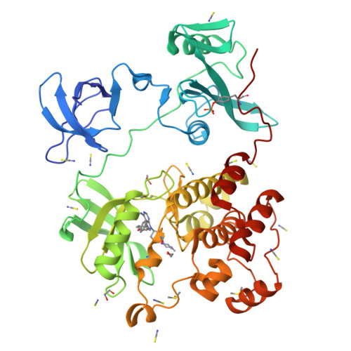
    Hematopoietic cell kinase (Hck) is a member of the Src kinase family and is a promising drug target in myeloid leukemias. Here, we report the crystal structure of human Hck in complex with the pyrrolopyrimidine inhibitor A-419259, determined at a resolution of 1.8 Å. This structure reveals the complete Hck active site in the presence of A-419259, including the αC-helix, the DFG motif, and the activation loop. A-419259 binds at the ATP-site of Hck and induces an overall closed conformation of the kinase with the regulatory SH3 and SH2 domains bound intramolecularly to their respective internal ligands. A-419259 stabilizes the DFG-in/αC-helix-out conformation observed previously with Hck and the pyrazolopyrimidine inhibitor PP1 (PDB: 1QCF). However, the activation loop conformations are distinct, with PP1 inducing a folded loop structure with the tyrosine autophosphorylation site (Tyr416) pointing into the ATP binding site, while A-419259 stabilizes an extended loop conformation with Tyr416 facing out into the solvent. Autophosphorylation also induces activation loop extension and significantly reduces the Hck sensitivity to PP1 but not A-419259. In cancer cells where Hck is constitutively active, the extended autophosphorylation loop may render Hck more sensitive to inhibitors like A-419259 which prefer this kinase conformation. More generally, these results provide additional insight into targeted kinase inhibitor design and how conformational preferences of inhibitors may impact selectivity and potency.


  • Organizational Affiliation

    Department of Microbiology and Molecular Genetics, University of Pittsburgh School of Medicine, 450 Technology Drive, Pittsburgh, Pennsylvania PA 15219, United States.


Macromolecules
Find similar proteins by:  (by identity cutoff)  |  3D Structure
Entity ID: 1
MoleculeChains Sequence LengthOrganismDetailsImage
Tyrosine-protein kinase HCK454Homo sapiensMutation(s): 3 
Gene Names: HCK
EC: 2.7.10.2
UniProt & NIH Common Fund Data Resources
Find proteins for P08631 (Homo sapiens)
Explore P08631 
Go to UniProtKB:  P08631
PHAROS:  P08631
GTEx:  ENSG00000101336 
Entity Groups  
Sequence Clusters30% Identity50% Identity70% Identity90% Identity95% Identity100% Identity
UniProt GroupP08631
Sequence Annotations
Expand
  • Reference Sequence
Small Molecules
Ligands 4 Unique
IDChains Name / Formula / InChI Key2D Diagram3D Interactions
VSE (Subject of Investigation/LOI)
Query on VSE

Download Ideal Coordinates CCD File 
B [auth A]7-[trans-4-(4-methylpiperazin-1-yl)cyclohexyl]-5-(4-phenoxyphenyl)-7H-pyrrolo[2,3-d]pyrimidin-4-amine
C29 H34 N6 O
FDVSOQRNTAPCHB-YHBQERECSA-N
DMS
Query on DMS

Download Ideal Coordinates CCD File 
D [auth A],
W [auth A]
DIMETHYL SULFOXIDE
C2 H6 O S
IAZDPXIOMUYVGZ-UHFFFAOYSA-N
EDO
Query on EDO

Download Ideal Coordinates CCD File 
C [auth A]
H [auth A]
I [auth A]
O [auth A]
X [auth A]
1,2-ETHANEDIOL
C2 H6 O2
LYCAIKOWRPUZTN-UHFFFAOYSA-N
SCN
Query on SCN

Download Ideal Coordinates CCD File 
E [auth A]
F [auth A]
G [auth A]
J [auth A]
K [auth A]
THIOCYANATE ION
C N S
ZMZDMBWJUHKJPS-UHFFFAOYSA-M
Modified Residues  1 Unique
IDChains TypeFormula2D DiagramParent
PTR
Query on PTR
A
L-PEPTIDE LINKINGC9 H12 N O6 PTYR
Experimental Data & Validation

Experimental Data

  • Method: X-RAY DIFFRACTION
  • Resolution: 1.80 Å
  • R-Value Free:  0.219 (Depositor), 0.219 (DCC) 
  • R-Value Work:  0.199 (Depositor), 0.199 (DCC) 
  • R-Value Observed: 0.200 (Depositor) 
Space Group: P 21 21 21
Unit Cell:
Length ( Å )Angle ( ˚ )
a = 43.346α = 90
b = 85.025β = 90
c = 128.798γ = 90
Software Package:
Software NamePurpose
PHENIXrefinement
Blu-Icedata collection
XDSdata reduction
PHASERphasing
Cootmodel building
XSCALEdata scaling

Structure Validation

View Full Validation Report


Ligand Structure Quality Assessment 

Created with Raphaël 2.3.0Worse 01 BetterLigand structure goodness of fit to experimental dataBest fitted VSEClick on this verticalbar to view details

Entry History & Funding Information

Deposition Data


Funding OrganizationLocationGrant Number
National Institutes of Health/National Cancer Institute (NIH/NCI)United StatesR01 CA233576
National Institutes of Health/National Cancer Institute (NIH/NCI)United StatesF31 CA265294

Revision History  (Full details and data files)

  • Version 1.0: 2024-10-09
    Type: Initial release
  • Version 1.1: 2024-10-30
    Changes: Database references