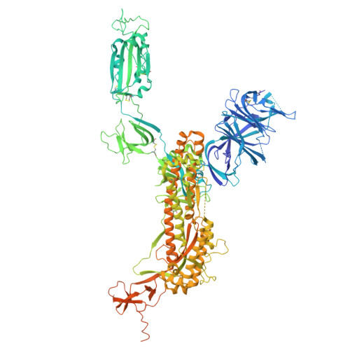
9C44

SARS-CoV-2 S + S2L20


Experimental Data Snapshot

  • Method: ELECTRON MICROSCOPY
  • Resolution: 2.60 Å
  • Aggregation State: PARTICLE 
  • Reconstruction Method: SINGLE PARTICLE 

wwPDB Validation   3D Report Full Report


This is version 1.0 of the entry. See complete history


Literature

Design of customized coronavirus receptors.

Liu, P.Huang, M.L.Guo, H.McCallum, M.Si, J.Y.Chen, Y.M.Wang, C.L.Yu, X.Shi, L.L.Xiong, Q.Ma, C.B.Bowen, J.E.Tong, F.Liu, C.Sun, Y.H.Yang, X.Chen, J.Guo, M.Li, J.Corti, D.Veesler, D.Shi, Z.L.Yan, H.

(2024) Nature 

  • DOI: https://doi.org/10.1038/s41586-024-08121-5
  • Primary Citation of Related Structures:  
    9C44, 9C45

  • PubMed Abstract: 

    Although coronaviruses use diverse receptors, the characterization of coronaviruses with unknown receptors has been impeded by a lack of infection models 1,2 . Here we introduce a strategy to engineer functional customized viral receptors (CVRs). The modular design relies on building artificial receptor scaffolds comprising various modules and generating specific virus-binding domains. We identify key factors for CVRs to functionally mimic native receptors by facilitating spike proteolytic cleavage, membrane fusion, pseudovirus entry and propagation for various coronaviruses. We delineate functional SARS-CoV-2 spike receptor-binding sites for CVR design and reveal the mechanism of cell entry promoted by the N-terminal domain-targeting S2L20-CVR. We generated CVR-expressing cells for 12 representative coronaviruses from 6 subgenera, most of which lack known receptors, and show that a pan-sarbecovirus CVR supports propagation of a propagation-competent HKU3 pseudovirus and of authentic RsHuB2019A 3 . Using an HKU5-specific CVR, we successfully rescued wild-type and ZsGreen-HiBiT-incorporated HKU5-1 (LMH03f) and isolated a HKU5 strain from bat samples. Our study demonstrates the potential of the CVR strategy for establishing native receptor-independent infection models, providing a tool for studying viruses that lack known susceptible target cells.


  • Organizational Affiliation

    State Key Laboratory of Virology, College of Life Sciences, TaiKang Center for Life and Medical Sciences, Wuhan University, Wuhan, China.


Macromolecules
Find similar proteins by:  (by identity cutoff)  |  3D Structure
Entity ID: 1
MoleculeChains Sequence LengthOrganismDetailsImage
Spike glycoproteinA,
D [auth B],
G [auth C]
1,288Severe acute respiratory syndrome coronavirus 2Mutation(s): 9 
Gene Names: S2
UniProt
Find proteins for P0DTC2 (Severe acute respiratory syndrome coronavirus 2)
Explore P0DTC2 
Go to UniProtKB:  P0DTC2
Entity Groups  
Sequence Clusters30% Identity50% Identity70% Identity90% Identity95% Identity100% Identity
UniProt GroupP0DTC2
Glycosylation
Glycosylation Sites: 15Go to GlyGen: P0DTC2-1
Sequence Annotations
Expand
  • Reference Sequence
Find similar proteins by:  (by identity cutoff)  |  3D Structure
Entity ID: 2
MoleculeChains Sequence LengthOrganismDetailsImage
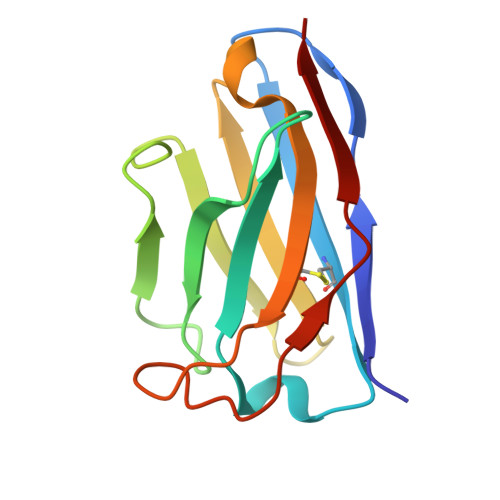
S2L20 Fab Heavy Chain VariableB [auth H],
E,
H [auth F]
121Homo sapiensMutation(s): 0 
Gene Names: S2
Entity Groups  
Sequence Clusters30% Identity50% Identity70% Identity90% Identity95% Identity100% Identity
Sequence Annotations
Expand
  • Reference Sequence
Find similar proteins by:  (by identity cutoff)  |  3D Structure
Entity ID: 3
MoleculeChains Sequence LengthOrganismDetailsImage
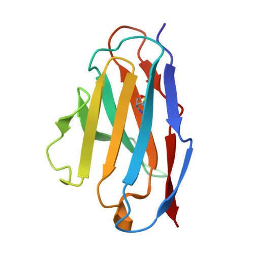
S2L20 Fab Light Chain Variable RegionC [auth L],
F [auth D],
I [auth G]
107Homo sapiensMutation(s): 0 
Gene Names: S2
Entity Groups  
Sequence Clusters30% Identity50% Identity70% Identity90% Identity95% Identity100% Identity
Sequence Annotations
Expand
  • Reference Sequence
Oligosaccharides

Help

Entity ID: 4
MoleculeChains Length2D Diagram Glycosylation3D Interactions
2-acetamido-2-deoxy-beta-D-glucopyranose-(1-4)-2-acetamido-2-deoxy-beta-D-glucopyranose
J [auth I],
L [auth K],
M,
N,
P,
J [auth I],
L [auth K],
M,
N,
P,
Q,
R,
T,
U
2N-Glycosylation
Entity ID: 5
MoleculeChains Length2D Diagram Glycosylation3D Interactions
beta-D-mannopyranose-(1-4)-2-acetamido-2-deoxy-beta-D-glucopyranose-(1-4)-2-acetamido-2-deoxy-beta-D-glucopyranoseK [auth J],
O,
S
3N-Glycosylation
Small Molecules
Ligands 1 Unique
IDChains Name / Formula / InChI Key2D Diagram3D Interactions
NAG
Query on NAG

Download Ideal Coordinates CCD File 
AA [auth A]
AB [auth C]
BA [auth A]
BB [auth C]
CA [auth A]
AA [auth A],
AB [auth C],
BA [auth A],
BB [auth C],
CA [auth A],
DA [auth A],
EA [auth A],
FA [auth A],
GA [auth B],
HA [auth B],
IA [auth B],
JA [auth B],
KA [auth B],
LA [auth B],
MA [auth B],
NA [auth B],
OA [auth B],
PA [auth B],
QA [auth B],
RA [auth C],
SA [auth C],
TA [auth C],
UA [auth C],
V [auth A],
VA [auth C],
W [auth A],
WA [auth C],
X [auth A],
XA [auth C],
Y [auth A],
YA [auth C],
Z [auth A],
ZA [auth C]
2-acetamido-2-deoxy-beta-D-glucopyranose
C8 H15 N O6
OVRNDRQMDRJTHS-FMDGEEDCSA-N
Experimental Data & Validation

Experimental Data

  • Method: ELECTRON MICROSCOPY
  • Resolution: 2.60 Å
  • Aggregation State: PARTICLE 
  • Reconstruction Method: SINGLE PARTICLE 

Structure Validation

View Full Validation Report


Entry History & Funding Information

Deposition Data


Funding OrganizationLocationGrant Number
Howard Hughes Medical Institute (HHMI)United States--

Revision History  (Full details and data files)

  • Version 1.0: 2024-11-20
    Type: Initial release