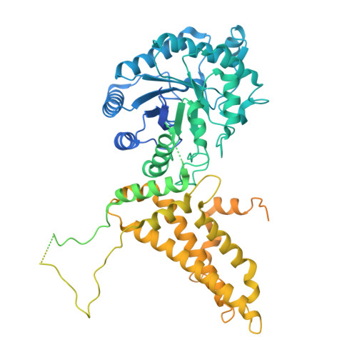
Human O-GlcNAcase catalytic-stalk dimer anchors flexible histone binding domains.
Nyenhuis, S.B., Steenackers, A., Mukherjee, M.M., Hinshaw, J.E., Hanover, J.A.(2025) Commun Chem 9: 8-8
- PubMed: 41366547
- DOI: https://doi.org/10.1038/s42004-025-01813-7
- Primary Citation of Related Structures:
9NE2, 9NE4, 9NE5 - PubMed Abstract:
Although thousands of proteins are specifically O-GlcNAc modified, the molecular features recognized by the enzymes of O-GlcNAc cycling (OGT/OGA) remain poorly defined. Here we solved the structure of the long isoform of human OGA (OGA-L) by cryo-electron microscopy (cryo-EM) providing a physiologically relevant platform to study the enzyme. The catalytic-stalk dimer structure was solved to a resolution of 3.63 Å, and the locally refined OGA A- and B-chains to 2.98 Å and 3.05 Å respectively. Intriguingly, the cryo-EM structures also exhibit lower resolution densities associated with the pHAT domains, suggesting substantial flexion of these domains relative to the catalytic-stalk dimer. OGA-L binds to a small subset of the 384 modified histone tails on a commercial histone peptide array. High affinity binding of OGA-L was detected to recombinant DNA-containing mononucleosomes bearing the H3K36 Me3 and H4K 5,8,12,16Ac modifications. The OGA-L-H3K36 Me3 interaction was further validated by traditional ChIP experiments in MEFs. Thus, OGA-L binds to two modified histone tails of nucleosomes linked to open chromatin, whereas it does not bind to marks associated with repressive chromatin. This model is consistent with OGA-L acting as a 'reader' of histone modifications linked to development, transcriptional activation, transposon silencing, and DNA damage repair.
- Laboratory of Molecular Biology, NIDDK, National Institutes of Health, Bethesda, MD, USA.
Organizational Affiliation: