9UEO | pdb_00009ueo

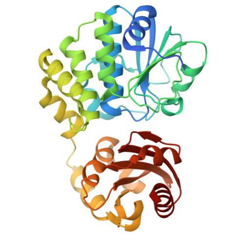
Co-crystal structure of Mtb CdnP with arabinose-derived 2'3'-cGAMP (AR-cGAMP) analogue


Experimental Data Snapshot

  • Method: X-RAY DIFFRACTION
  • Resolution: 2.60 Å
  • R-Value Free: 
    0.250 (Depositor), 0.260 (DCC) 
  • R-Value Work: 
    0.210 (Depositor), 0.218 (DCC) 

Starting Model: experimental
View more details

wwPDB Validation 3D Report Full Report

Validation slider image for 9UEO

Ligand Structure Quality Assessment 


This is version 1.0 of the entry. See complete history

Literature

Structural and Biochemical Insights into the Arabinose and Xylose Derivatives of Cyclic-GAMP-Mediated Inhibition of Mycobacterium tuberculosis Cyclic-di-AMP Phosphodiesterase.

Hanuman, D.S.Neeharika, S.Nitin, K.Abhishek, S.Sinha, K.M.Rajakumara, E.

(2026) Biochemistry 65: 2441-2451

  • DOI: https://doi.org/10.1021/acs.biochem.6c00106
  • Primary Citation Related Structures: 
    9UEO

  • PubMed Abstract: 

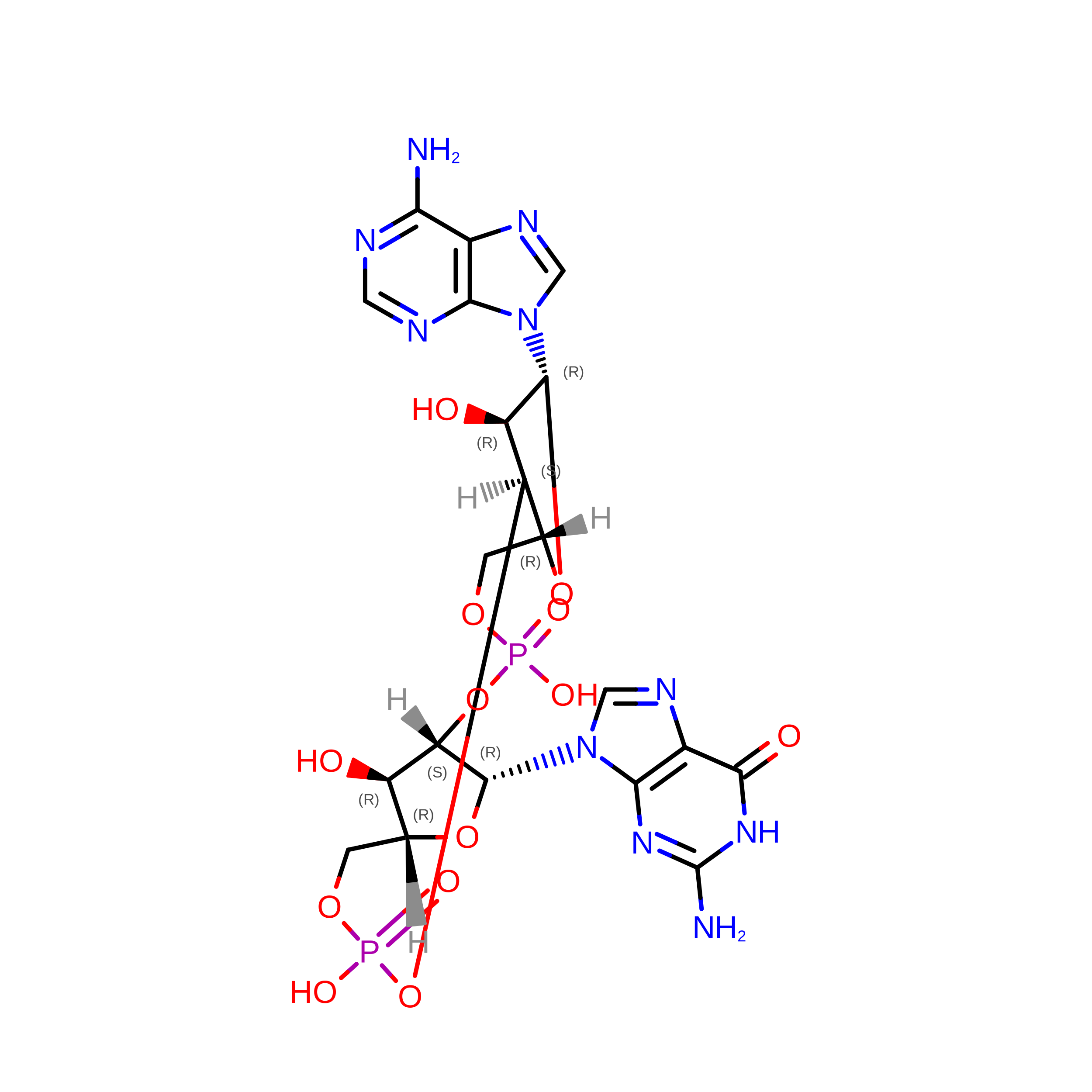
    Bacterial infection, including that ofMycobacterium tuberculosis, leads to the accumulation of the bacterial cyclic dinucleotides (CDN), c-di-AMP and c-di-GMP, and the host cGAMP synthase-catalyzed CDN, 2'3'-cGAMP in the cytosol, which activates STING-dependent type I interferon (IFN) and NF-κB immune responses. The mycobacterial cyclic dinucleotide phosphodiesterase (CdnP) secreted into the host macrophages blunts host immunity by directly cleaving bacterial- and host-derived CDNs. The arabinose- and xylose-modified 2'3'-cGAMP (2'3'-(A/X)cGAMP) analogues act as potent STING agonists and resist hydrolysis by the host-PDE ENPP1. Here, we report that 2'3'-(A/X)-cGAMP analogues bind to CdnP and compete with its substrate binding. Further studies revealed that these analogues inhibit the catalytic activity of CdnP. The cocrystal structure demonstrates that the arabinose-derived 2'3'-cGAMP (AR-cGAMP) analogue is trapped in an unusual U-shaped conformation in the substrate-binding pocket, away from the catalytic residue and Mn2+, which suggests that CdnP is incompetent to hydrolyze the analogue and cannot accept the other CDN substrate for hydrolysis. Given that several bacterial and viral pathogens deploy CDN phosphodiesterase enzymes to hydrolyze both host and pathogen-derived STING agonists, sugar-modified CDNs can be used to weaken bacterial and viral defenses and stimulate the STING-mediated host immunity against these pathogens.


  • Organizational Affiliation
    • Macromolecular Structural Biology Laboratory, Department of Biotechnology, Indian Institute of Technology Hyderabad (IITH), Hyderabad, Telangana502285, India.

Macromolecule Content 

  • Total Structure Weight: 108.62 kDa 
  • Atom Count: 7,145 
  • Modeled Residue Count: 954 
  • Deposited Residue Count: 1,008 
  • Unique protein chains: 1

Macromolecules

Find similar proteins by:|  3D Structure
Entity ID: 1
MoleculeChains  Sequence LengthOrganismDetailsImage
Bifunctional oligoribonuclease and PAP phosphatase NrnA
A, B, C
336Mycobacterium tuberculosis H37RvMutation(s): 0 
Gene Names: nrnARv2837c
EC: 3.1 (PDB Primary Data), 3.1.3.7 (PDB Primary Data)
UniProt
Find proteins for P71615 (Mycobacterium tuberculosis (strain ATCC 25618 / H37Rv))
Explore P71615 
Go to UniProtKB:  P71615
Entity Groups
Sequence Clusters30% Identity50% Identity70% Identity90% Identity95% Identity100% Identity
UniProt GroupP71615
Sequence Annotations
Expand
Reference Sequence

Small Molecules

Ligands 3 Unique
IDChains Name / Formula / InChI Key2D Diagram3D Interactions
9UH
(Subject of Investigation/LOI)

Query on 9UH


Download:Ideal Coordinates CCD File
D [auth A],
G [auth B],
I [auth C]
(2R,5R,7R,8S,10R,12aR,14R,15R,15aS,16R)-7-(2-amino-6-oxo-1,6-dihydro-9H-purin-9-yl)-14-(6-amino-9H-purin-9-yl)-2,10,15,16-tetrahydroxyoctahydro-2H,10H,12H-5,8-methano-2lambda~5~,10lambda~5~-furo[3,2-l][1,3,6,9,11,2,10]pentaoxadiphosphacyclotetradecine-2,10-dione
C20 H24 N10 O13 P2
XRILCFTWUCUKJR-SOFRMEQTSA-N
EDO

Query on EDO


Download:Ideal Coordinates CCD File
F [auth A]1,2-ETHANEDIOL
C2 H6 O2
LYCAIKOWRPUZTN-UHFFFAOYSA-N
MN

Query on MN


Download:Ideal Coordinates CCD File
E [auth A],
H [auth B],
J [auth C]
MANGANESE (II) ION
Mn
WAEMQWOKJMHJLA-UHFFFAOYSA-N

Experimental Data & Validation

Experimental Data

  • Method: X-RAY DIFFRACTION
  • Resolution: 2.60 Å
  • R-Value Free:  0.250 (Depositor), 0.260 (DCC) 
  • R-Value Work:  0.210 (Depositor), 0.218 (DCC) 
Space Group: I 21 21 21
Unit Cell:
Length ( Å )Angle ( ˚ )
a = 78.4α = 90
b = 148.92β = 90
c = 159.4γ = 90
Software Package:
Software NamePurpose
PHENIXrefinement
Aimlessdata scaling
iMOSFLMdata reduction
REFMACrefinement
MOLREPphasing

Structure Validation

View Full Validation Report


Ligand Structure Quality Assessment 


Entry History 

& Funding Information

Deposition Data


Funding OrganizationLocationGrant Number
Not funded--

Revision History  (Full details and data files)

  • Version 1.0: 2026-09-16
    Type: Initial release