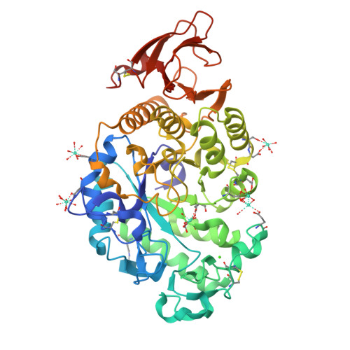
Crystal Structure of alpha-amylase from Aspergillus oryzae
Sugahara, M.To be published.
Experimental Data Snapshot
Entity ID: 1 | |||||
|---|---|---|---|---|---|
| Molecule | Chains | Sequence Length | Organism | Details | Image |
| Alpha-amylase A type-1/2 | 478 | Aspergillus oryzae RIB40 | Mutation(s): 0 EC: 3.2.1.1 | 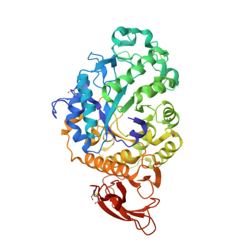 | |
UniProt | |||||
Find proteins for P0C1B3 (Aspergillus oryzae (strain ATCC 42149 / RIB 40)) Explore P0C1B3 Go to UniProtKB: P0C1B3 | |||||
Entity Groups | |||||
| Sequence Clusters | 30% Identity50% Identity70% Identity90% Identity95% Identity100% Identity | ||||
| UniProt Group | P0C1B3 | ||||
Glycosylation | |||||
| Glycosylation Sites: 1 | |||||
Sequence AnnotationsExpand | |||||
| |||||
| Ligands 3 Unique | |||||
|---|---|---|---|---|---|
| ID | Chains | Name / Formula / InChI Key | 2D Diagram | 3D Interactions | |
| NAG Query on NAG | B [auth A] | 2-acetamido-2-deoxy-beta-D-glucopyranose C8 H15 N O6 OVRNDRQMDRJTHS-FMDGEEDCSA-N |  | ||
| GD Query on GD | C [auth A] D [auth A] E [auth A] F [auth A] G [auth A] | GADOLINIUM ATOM Gd UIWYJDYFSGRHKR-UHFFFAOYSA-N |  | ||
| CA Query on CA | I [auth A] | CALCIUM ION Ca BHPQYMZQTOCNFJ-UHFFFAOYSA-N |  | ||
| Length ( Å ) | Angle ( ˚ ) |
|---|---|
| a = 48.524 | α = 90 |
| b = 65.619 | β = 90 |
| c = 130.243 | γ = 90 |
| Software Name | Purpose |
|---|---|
| HKL-2000 | data collection |
| CNS | refinement |
| HKL-2000 | data reduction |
| HKL-2000 | data scaling |
| CNS | phasing |